3316
Blood flow MRI of the mouse optic nerve head1Radiology, Stony Brook University, Stony Brook, NY, United States
Synopsis
Different retinal diseases likely affect the retinal, choroidal, and optic nerve head (ONH) circulations differently. Methods to quantitatively measure ONH blood flow with depth resolution are lacking. In this study, a blood flow MRI method was optimized to image blood flow of the ONH, retina, and choroid in the mouse eye at 42x42x275µm. This method could be useful to study models of retinal disease, such as glaucoma.
Introduction
Different retinal diseases likely affect the retinal, choroidal, and optic nerve head (ONH) circulations differently. The retina has two separate blood supplies with substantial differences, the retinal and the choroidal vessels, with choroidal blood flow (BF) being many times greater than retinal BF. The optic nerve head (ONH) has vascular supplies arising from the retinal circulation, the short posterior ciliary arteries (which also supply the choroid), and the central retinal artery. In glaucoma, elevated intraocular pressure can deform the optic nerve head, which may pinch the vessels, impairing blood flow. Thus, localized blood flow measurement at the optic nerve head may be important in glaucoma. The ability of MRI to distinguish retinal BF and choroidal BF has previously been demonstrated (1), but techniques to quantify the ONH BF are lacking. The goal of this study was to further improve the resolution of BF MRI of the mouse eye to 42x42x275µm to distinguish the optic nerve head BF.Methods
Blood flow of the optic nerve head, retina, and choroid was measured using arterial spin labeling (ASL) MRI on a 7T/30cm scanner (Bruker) with a 1500 mT/m gradient. C57BL/6 mice (n=3, female) were anesthetized with 1.5% isoflurane. Temperature (~37oC) and respiration rate (~90 breaths/min) were monitored and maintained. A small surface eye coil (diameter=6 mm) was used for imaging. Continuous ASL was performed with a circular coil (diameter=8 mm) for labeling placed at the heart (1,2). Images were acquired with a single slice bisecting the eye with gradient-echo EPI with FOV=6x6 mm, matrix=144x144, 3 segments, 275 µm slice thickness, TR=3.0s, TE=7.9ms, labeling duration=2.1s, and post labeling delay=300ms. BF was calculated as in (3), and the retina was flattened for profile analysis (1) to analyze the retinal, choroidal, and ONH BF.Results
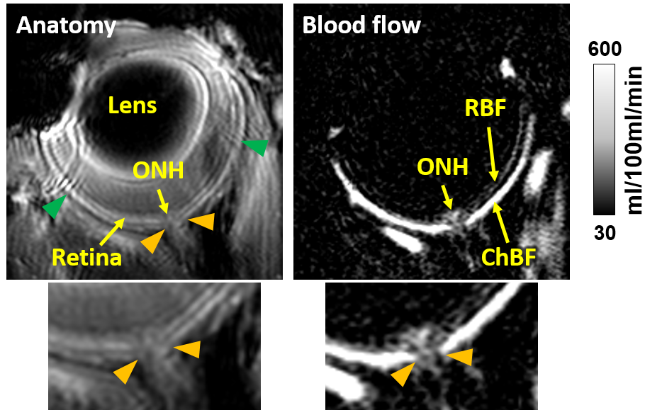
An example ocular blood flow map is shown in Figure 1. At the area where the optic nerve penetrates through the retina, the anatomical layer structures of the retina are absent as expected, and similarly the bright choroidal BF layer is absent at the ONH on the BF map. To analyze retinal and choroidal BF, profiles along the retina on both sides of the ONH were averaged (Figure 1), and to analyze ONH BF profiles along the ONH region were averaged (Figure 1). Group-average profiles of the BF of the retina/choroid and of the ONH are shown in Figure 2. ONH BF (averaged over a 299µm region from the vitreous-retinal boundary to the choroid-sclera boundary) was 278±22.6 ml/100ml/min (mean±SEM), relatively higher than the retinal BF of 141±13 ml/100ml/min but lower than choroidal BF of 637±67 ml/100ml/min.Discussion
This study demonstrates a novel approach to quantitatively image the ONH BF using MRI. BF of the retinal and choroidal circulations can also be quantified using this method. Choroidal BF was many times larger than retinal BF, as previously reported (1). A few previous studies have used the microsphere method to measure ONH BF in primates, but microsphere measurements are highly invasive and are terminal as the tissue must be analyzed post-mortem. These primate studies looking at ONH BF as a function of depth from the surface found similar results as ours, in that ONH BF is relatively high in the regions at the level of the retina/choroid, but then rapidly drops to quite low going behind the sclera (4,5). In primates the retinal BF was reported to be about 120-150g/100ml/min and the ONH BF (at the depth of the retina/choroid) was reported to be about 150-250 g/100ml/min (4), in reasonable agreement with our MRI results in mice.Methods to quantitatively measure ONH BF with depth resolution are lacking. Our MRI approach can provide important BF data that is not depth limited and is non-invasive. This method could be useful to study models of retinal disease, such as glaucoma.
Acknowledgements
This work was supported by NIH R01 EY027751 (TQD).References
1. Muir ER, Duong TQ. MRI of retinal and choroidal blood flow with laminar resolution. NMR Biomed 2011;24:216-223.
2. Muir ER, Shen Q, Duong TQ. Cerebral blood flow MRI in mice using the cardiac spin-labeling technique. Magn Reson Med 2008;60:744-748.
3. Peng Q, Zhang Y, Nateras OS, van Osch MJ, Duong TQ. MRI of blood flow of the human retina. Magn Reson Med 2011;65:1768-1775.
4. Sperber GO, Bill A. Blood flow and glucose consumption in the optic nerve, retina and brain: effects of high intraocular pressure. Exp Eye Res 1985;41:639-653.
5. Geijer C, Bill A. Effects of raised intraocular pressure on retinal, prelaminar, laminar, and retrolaminar optic nerve blood flow in monkeys. Invest Ophthalmol Vis Sci 1979;18:1030-1042.
Figures