3305
Experimental comparison of velocity selective ASL schemes1FMRI Laboratory, University of Michigan, Ann Arbor, MI, United States, 2University of California at Riverside, Riverside, CA, United States
Synopsis
This work presents an experimental comparison of different velocity selective (VS) labeling strategies for non-contrast perfusion MRI, including new variants of VS saturation and VS inversion, in relation to pseudo-continuous labeling pulses (PCASL).
Introduction
Arterial Spin Labeling techniques allow us to image brain perfusion without the use of contrast injections. While the standard labeling technique is currently a train of pseudo-continuous pulses (PCASL) [Dai 2008], velocity selective labeling pulses are a very attractive alternative because of their insensitivity to arterial bolus arrival time, which can be highly variable in neurological disease. Multiple approaches and pulses for velocity selective labeling have been proposed with different velocity profiles and robustness to imperfect B1 and B0 fields.This work focuses on comparing different velocity selective (VS) labeling pulses for non-contrast perfusion MRI, including new variants of VS saturation and inversion pulses in relation to pseudo-continuous labeling pulses (PCASL). Specifically, the aims are to evaluate the pulses in terms of their velocity profiles , robustness to B1 and B0 imperfections, magnitude of perfusion signal, and temporal SNR in vivo.
Methods
Participants were imaged using a 3T MR750 scanner (General Electric, Waukesha, WI) in compliance to our institution’s internal review board’s regulations. The read-out in all experiments was the same: a custom 3D fast spin echo, stack-of-spirals sequence with echo time of 15 ms. and 18 echoes. (TR=4600ms, matrix=64x63, 18 slices, BW = 85 kHz). Each echo corresponded to a different kz encoding step. A spiral encoding readout trajectory with two interleaves was used.In all subjects, twelve pairs of images were collected in each case. The first pair in the series was collected without any labeling pulses and was used as the reference image (M0).
We tested the following labeling schemes in six participants (1) PCASL (2) “VSI-sinc” and (3) “VSI-sinc 2”: two variants of the FTVSI pulse based on a sinc pulses and composite refocusing pulses as in [Landes 2019] (4) “VSI-rect”: original Fourier Transform VS inversion pulses based on a rect pulse [Qin 2016](5) “VSS”: a single VS saturation pulses using a symmetric BIR-8 [Guo 2014]. (6) “VSSx2”: two consecutive such saturation pulses [Guo 2015] (3) “VSS Inv”: a single saturation pulse using a modified BIR-8 pulse that tips the spins to the negative z axis in the last segment [Guo 2015] .
The VS labeling pulses were applied 1300 ms prior to readout, except in the VSSx2 case (TI1=1150ms, TI2=820ms). No background suppression (BGS) pulses were used in any of the sequences.
All VSASL were collected with a cutoff velocity of 2 cm/s and an arterial suppression pulse to cotrol the bolus duration, consisting of a sym-BIR-8 train with the same cutoff velocity, applied 100 ms before acquisition. PCASL images were also collected in both experiments as reference. An 1800 ms labeling period was used, followed by a 2000 ms labeling delay to avoid arterial vascular artefacts.
Percent signal change and temporal signal-to-noise ratio (tSNR) were computed on a voxelwise basis. The tSNR was computed by dividing the mean signal change over the standard deviation in time. The percent signal change and tSNR were averaged over each subject’s brain. PCASL data was excluded in one subject and VSSx2 was excluded on another subject because of experimental error.
Results
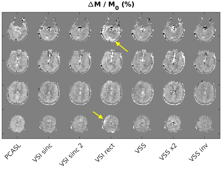
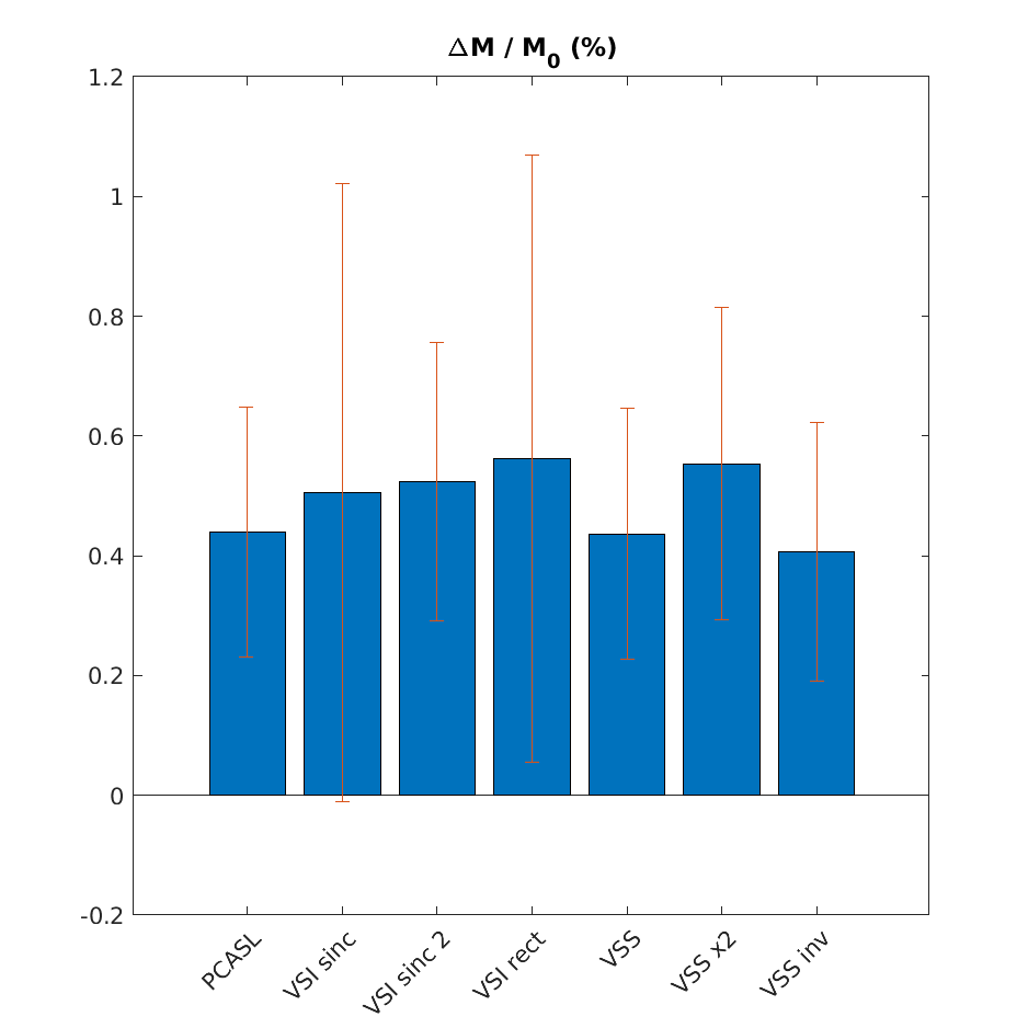
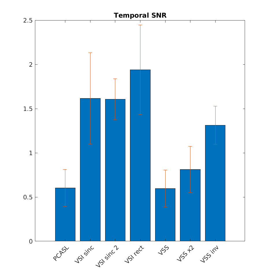
Figure 1 shows the percent signal difference between control (non-selective) and label (selective) images collected with the inversion labeling schemes in a representative subject. The original rect VSI pulse can produce noticeable artefacts relative to the sinc-based versions. The windowed sinc pulse (“sinc 2”) show the least amount of artefact.Figure 2 shows a summary of the average percent signal change per voxel for each of the labeling schemes under scrutiny. Similarly, figure 3 shows a summary of their temporal SNR. The bar graphs indicate the mean and std. dev. across subjects. The figure indicates that the original pulse by Qin et al has the highest SNR, but its performance is the most variable from subject to subject and yields artefacts (see fig 1 ) due to sensitivity to B1 or B0 inhomogeneity
Discussion
Our experimental results are in agreement with theoretical predictions and numerical simulations (submitted in a companion abstract for this conference). The FT-VSI pulse based on a windowed sinc seems to provide the best compromise in terms of contrast and stability.VSI-sinc pulses produce a sharper (no ripples) velocity profile than VSI-rect pulses in simulation and yield a more stable signal in vivo. However, VSI pulses still only yield a modest improvement in temporal SNR relative to VS saturation schemes. VS saturation pulses with inversion produce similar results. The dual saturation scheme produces the greatest contrast, although not the highest temporal SNR, which could be improved by additional BGS pulses.
The Inversion of stationary spins in VSI and VSS-inv pulses results in partial suppression of the background signal, and thus reduces the variance of the ASL subtraction.
Acknowledgements
No acknowledgement found.References
Dai WY, Garcia D, de Bazelaire C, et al. Continuous Flow-Driven Inversion for Arterial Spin Labeling Using Pulsed Radio Frequency and Gradient Fields. Magn Reson Med 2008; 60: 1488-1497.
Guo J, Meakin JA, Jezzard P, et al. An optimized design to reduce eddy current sensitivity in velocity-selective arterial spin labeling using symmetric BIR-8 pulses. Magn Reson Med 2014; 73: 1085-1094.
Guo J and Wong EC. Increased SNR efficiency in velocity selective arterial spin labeling using multiple velocity selective saturation modules (mm-VSASL). Magn Reson Med 2015; 74: 694-705.
Landes V, Jao TR, Javed A, Nayak KS . Improved velocity-selective labeling pulses for myocardial ASL. Proc. ISMRM 2019, num. 4967
Qin Q, van Zijl PCM. Velocity-selective-inversion prepared arterial spin labeling. Magn Reson Med. 2016;76(4):1136-1148. doi:10.1002/mrm.26010
Figures