3230
Multiple dual-echo reconstructions to improve the robustness of water-fat MRI
Samir D. Sharma1
1Canon Medical Research USA, Mayfield Village, OH, United States
1Canon Medical Research USA, Mayfield Village, OH, United States
Synopsis
A challenge in water-fat imaging is robust separation of the water and fat components. In this work, we propose a method that performs a series of dual-echo reconstructions using different pairs of echoes. The estimates from the multiple reconstructions are then combined to generate a more robust initial estimate of the water and fat images. The final estimates are then calculated via voxel-wise minimization. The proposed multiple dual-echo method demonstrates an improvement to single dual-echo reconstruction for water-fat imaging.
PURPOSE
The purpose of this work is to demonstrate a method to improve the robustness of water-fat imagingINTRODUCTION
Multi-echo water-fat imaging methods are routinely used for separation and quantification of water and fat. A challenge in water-fat imaging is robust separation of the water and fat components. Inaccurate separation of the water and fat components may degrade the diagnostic utility of the respective images as well as subsequently calculated quantities like the proton density fat fraction (PDFF). Therefore, consistently accurate separation of water and fat is necessary to maximize diagnostic utility of the method.Water-fat imaging can be performed via a two-step approach. In the first step, initial estimates of the water and fat (and possibly the B0 field and R2*) components are calculated. The next step refines the initial estimates to generate final estimates. Many multi-echo reconstruction techniques exist to generate the initial estimates1-4.
Recently, Zhong et al. proposed a method that uses a dual-echo reconstruction to generate the initial estimates of water and fat5. Unlike multi-echo reconstructions, dual-echo reconstructions do not assume that the phase evolves linearly with time. Therefore, they are more robust than multi-echo reconstructions when the background phase does not evolve linearly. The dual-echo reconstruction5 uses only two echoes from the multi-echo acquisition. Information contained in the other echoes may be useful for a more consistent initial estimation of water and fat. However, this method ignores those echoes. In this work, we demonstrate a method to improve the robustness of water-fat imaging. The proposed method makes use of the multiple echoes by performing a series of dual-echo reconstruction using different pairs of echoes. The results are then combined to generate a more robust initial estimate of the water and fat images.
METHODS
Figure 1 demonstrates the proposed method. From a multi-echo acquisition, multiple different pairs of images are processed using a dual-echo reconstruction technique6-9. The water and fat images are estimated for each dual-echo reconstruction. The estimates are then combined across the different dual-echo reconstructions to generate an initial estimate of the water and fat images. The initial estimates are then refined using a voxel-wise minimization over all of the acquired echoes.To demonstrate this method, ten 3D axial liver datasets were acquired on a Canon Medical Titan 3T scanner under IRB approval. Acquisition parameters included: TE1=1.2 ms, ΔTE=1.0 ms, nTE=6, FOV=40x34, matrix=224x160, slice thickness=6 mm, number of slices=30, SPEEDER parallel imaging R=2, and scan time=19 seconds. Additionally, datasets from the ISMRM water-fat toolbox10 and the ISMRM water-fat reconstruction challenge11 were also processed.
For each dataset, both a single dual-echo reconstruction5 and the proposed multiple dual-echo reconstruction method were done. Dual-echo reconstruction was done using a flexible dual-echo technique8,9. For the single dual-echo reconstruction, the estimated water and fat images were used as the initial estimate for the subsequent voxel-wise minimization. For the multiple dual-echo reconstruction, the estimated water and fat images across all of the reconstructions were compared on a voxel-wise basis to decide the initial water and fat estimates for the subsequent voxel-wise minimization. Voxel-wise minimization was performed on the magnitude images to avoid phase errors caused by factors such as eddy currents. The voxel-wise minimization also included estimation of R2* to avoid confounding the PDFF estimate.
The final water and fat estimates were reviewed for the presence of water-fat swaps. If a swap existed, it was further classified as either a major swap or minor swap. A swap was considered to be major if it affected the anatomy of interest (e.g. liver) or corrupted at least 25% of the image, otherwise the swap was considered to be minor.
RESULTS
A summary of the image review is shown in Figure 2. In five cases, the multiple dual-echo method improved the swap classification versus the single dual-echo method. In two of the five cases, the classification improved from ‘major swap’ to ‘no swap’. In two other cases, the classification improved from ‘minor swap’ to ‘no swap’, and in one other case, the classification improved from ‘major swap’ to ‘minor swap’. In one case, the multi dual-echo reconstruction worsened the swap classification from ‘no swap’ to ‘minor swap’. All swaps occurred in either the ISMRM water-fat toolbox datasets10 or the ISMRM reconstruction challenge datasets11, which were created to be difficult datasets.Figure 3 shows an example case when the multiple dual-echo method improved the water-fat estimation in the abdomen. Figure 4 shows an example case when the multiple dual-echo method improved the water-fat estimation in the ankle.
DISCUSSION
The proposed multiple dual-echo method demonstrates an improvement to single dual-echo reconstruction for water-fat imaging. The multiple dual-echo method is less sensitive to swaps in any one single dual-echo reconstruction. These swaps could arise from an unfavorable choice of echo times or challenging anatomy.The benefit of the proposed method comes with an increased computational cost because multiple (three in this work) dual-echo reconstructions must be performed. In this work, the cost was an increased reconstruction time that was between 20-60 seconds (dependent on the dataset size). Parallelizing the multiple reconstructions should reduce the computation time, and is an item of potential future work.
Acknowledgements
The author thanks Bin Xie (Canon Medical China) for his assistance developing the reconstruction.References
- Yu H, Reeder SB, Shimakawa A, et al. Field map estimation with a region growing scheme for iterative 3‐point water‐fat decomposition. MRM 2005;54:1032-1039.
- Lu W and Hargreaves BA. Multiresolution field map estimation using golden section search for water‐fat separation. MRM 2008;60:236-244.
- Tsao J and Jiang Y. Hierarchical IDEAL: fast, robust, and multiresolution separation of multiple chemical species from multiple echo times. MRM 2013;70:155-159.
- Hernando D, Kellman P, Haldar JP, Liang ZP. Robust water/fat separation in the presence of large field inhomogeneities using a graph cut algorithm. MRM 2010;63:79-90.
- Zhong X, Nickel MD, Kannengiesser SA, et al. Liver fat quantification using a multi‐step adaptive fitting approach with multi‐echo GRE imaging. MRM 2014;72:1353-1365.
- Ma J. Breath‐hold water and fat imaging using a dual‐echo two‐point Dixon technique with an efficient and robust phase‐correction algorithm. MRM 2004;52:415-419.
- Xiang QS. Two‐point water‐fat imaging with partially‐opposed‐phase (POP) acquisition: an asymmetric Dixon method. MRM 2006;56:572-84.
- Berglund J, Ahlström H, Johansson L, Kullberg J. Two‐point dixon method with flexible echo times. MRM 2011;65:994-1004.
- Eggers H, Brendel B, Duijndam A, Herigault G. Dual‐echo Dixon imaging with flexible choice of echo times. MRM 2011;65:96-107.
- https://www.ismrm.org/workshops/FatWater12/data.htm
- https://challenge.ismrm.org/node/8
Figures

Figure
1:
Flowchart of
the proposed method. Example is shown for a commonly-used six-echo acquisition.
Multiple different pairs of dual-echo images are processed using a dual-echo
separation method. The estimates are pooled together to form an initial
estimate of water and fat. The initial estimates are then refined using all of
the acquired echoes.

Figure
2: Summary of swap analysis. The number in parenthesis is the number of cases
in which the water-fat swap classification for the multiple dual-echo
reconstruction was different than the single dual-echo reconstruction.
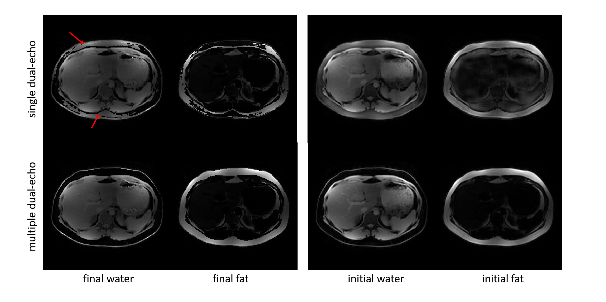
Figure
3: The proposed multiple dual-echo reconstruction results in water and fat
estimates without swap. The single dual-echo reconstruction contains swaps (red
arrows). Notice that the initial water and fat estimates in the single
dual-echo reconstruction suffer from incomplete separation. This error
propagates to the final estimates.
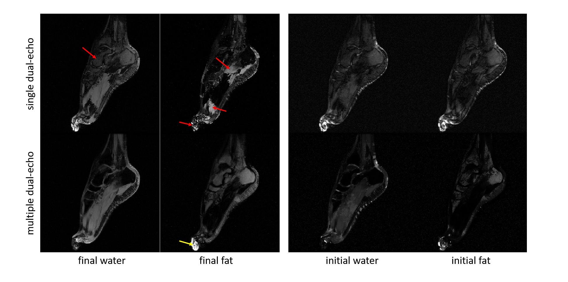
Figure
4: The proposed multiple dual-echo reconstruction results in water and fat
estimates with fewer swaps than the single dual-echo reconstruction. A swap
remains in the multi dual-echo reconstruction (yellow arrow). The single
dual-echo reconstruction contains many swaps (red arrows).