3188
Feasibility of Phase-based EPT using clinical routine sequences.1electrical electronic engineering, yonsei University, seoul, Korea, Republic of, 2Department of Radiology, Gangnam Severance Hospital, seoul, Korea, Republic of
Synopsis
In this study, we test whether the preparation pulse and compensation gradient affect B1-phase and suggested a method for merging B1-phase obtained from different pulse sequences. We also try to reconstruct conductivity with the routine of guide images for surgery which have high resolution and low SNR.
Introduction
Phase-based EPT(Electrical Property Tomography) is a technique to reconstruct tissue electrical property using only transceive phase information [1]. This technique has the advantage of no additional hardware installation and current injection. Therefore, the phase-based EPT method has the potential for immediate clinical usage. However, EPT method has an inherent noise amplification problem in the reconstruction process. To overcome the lack of SNR, fitting[2] and regularizing methods have been proposed.[3] But, these methods cause blurring effects on the resolution and contrast of reconstructed conductivity. To minimize the blurring effect on conductivity reconstruction, averaging the signal is advantageous to increase SNR. The phase-based EPT using TE=0 phase of the acquired image as B1-phase to calculate conductivity, i.e. Spin-Echo and bSSFP sequences phase. In clinical routine, inversion or spatial saturation pulse and flow compensation gradient are used depending on the application. In this study, we test whether the preparation pulse and compensation gradient affect B1-phase and suggested a method for merging B1-phase obtained from different pulse sequences. We also try to reconstruct conductivity with the routine of guide images for surgery which have high resolution and low SNR.Methods
1. Data Acquisition :Healthy volunteer - 3T clinical scanner (Skyra; Siemens Medical Solutions, Erlangen, Germany). Turbo Spin echo(TSE) with resolution = 1 × 1 mm2, FOV = 192 × 192 mm2, TR = 4500 ms, TEeff = 100ms, ETL = 15, slice thickness = 4 mm, number of slice =10, averaging =3. Acquire the same parameter for flow-compensation, spatial saturation. ~3min for each scan. FLAIR sequence with the same parameter except for repetition time = 6500ms, averaging =2. Scan time = 2min 50sec.
Meningioma Patient (cavernous malformation)– 3T clinical scanner (MR750, GE Healthcare, Waukesha, WI, USA). Turbo Spin echo(TSE) with resolution = 0.625 ×0.75mm2 , FOV= 240 ×240 mm2, slice thickness = 4 mm, TR =4363 ms, TEeff = 96ms, ETL = 15, number of slice =100, ~6min 30sec for each scan. FLAIR sequence with resolution = 0.5966 × 0.9375mm2, FOV = 210×210mm2, slice thickness = 4 mm, TI=2500ms, TR =10000 ms, TEeff =134ms, ETL = 28, number of slice =30, ~4min for each scan.
This study was approved by our Institutional Review Board (IRB).
2. Coil-combine method : a) TSE multi-coil images combined by using B1- based method and find coil coefficients.[4] b) applying coil-coefficients to other multi-coil images.
3. Registration & averaging: using mutual information TSE image registered to FLAIR image.[5] Registered TSE image with FLAIR image was averaged. To consider different SNR of images, Noise-normalized sum was used.
$$S_{normalized sum}=\frac{S_{TSE}(x)}{\sigma_{TSE}}+\frac{S_{FLAIR}(x)}{\sigma_{FLAIR}}$$
$$$(S_{TSE},S_{FLAIR}$$$ : signal of TSE and FLAIR, respectively.
And $$$\sigma_{TSE},\sigma_{FLAIR}$$$ are noise standard deviation of TSE and FLAIR, respectively. )
4. EPT reconstruction : A 2D-weighted polynomial fitting was performed to reconstruct conductivity [6]. (maximum kernel size = 1.9 x 1.9 cm2) For the magnitude weighting factor, the magnitude of the combined image was used.
Result
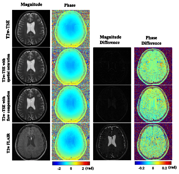
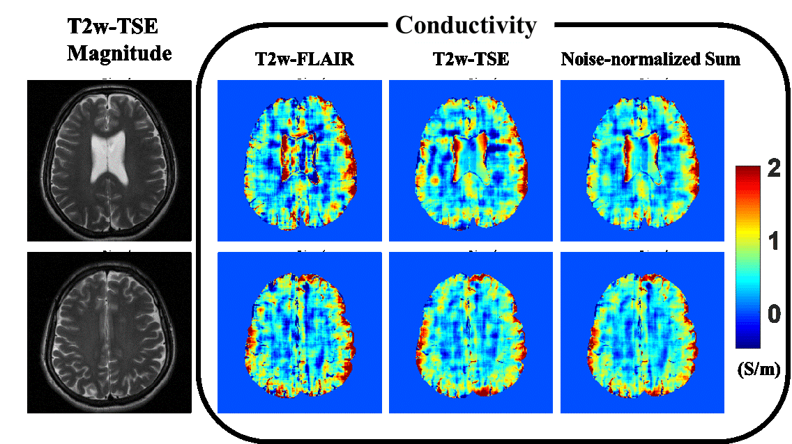
Figure 1 shows the magnitude and phase image of TSE and with spatial saturation, flow compensation and FLAIR. The difference map of magnitude and phase were presented to show the B1 phase is the same, whether with or without inversion pulse, flow compensation, and spatial saturation. As shown in the phase difference map, preparation pulse or compensation doesn’t affect B1-phase except for vein or CSF. SNR of FLAIR and TSE are 22.4 and 31.05, and SNR of Noise-normalized sum of them is 37.7.In Figure.2, shows the conductivity image of FLAIR, TSE and Noise-normalized sum. Fluctuation pattern of conductivity in white matter is reduced in Noise-normalized sum.
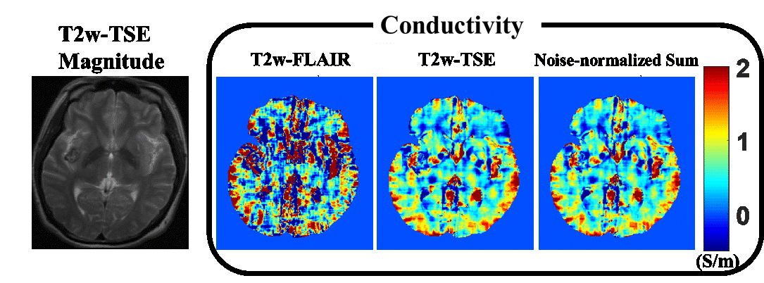
Figure 3 is a meningioma patient case. SNR of FLAIR was 13.2 and SNR of TSE after resizing and registration was 54.5. SNR of Noise-normalized sum was 56.4. SNR of averaged data only changed 2, because, SNR of FLAIR was too low. The conductivity result of averaged data has small differences with the conductivity reconstruction of TSE.
Discussion and Conclusion
To apply EPT reconstruction to clinical data, the trade-off between scan time and SNR could be a problem. In this study, the pipeline for averaging different scans with different sequences and FOV was introduced. But as shown in the patient case, the effect of these basic averaging strategies was limited. In future work, not only averaging before reconstruction but also conductivity combine method after conductivity reconstruction and how to utilize the segmentation information from different contrast images should be studied.Acknowledgements
No acknowledgement found.References
1. T. Voigt. et al., Quantitative conductivity and permittivity imaging of the human brain using electrical properties tomography. Mag. Reson. Med., Vol 66(2), August 2011, Pages 456-466.
2. Katscher U. et al., Determination of Electric Conductivity and Local SAR Via B1 Mapping. IEEE TMI., Vol 28(9), September 2009, Pages 1365-1373.
3. Marques JP. et al., Single acquisition electrical property mapping based on relative coil sensitivities: A proof-of-concept demonstration. Mag. Reson. Med., Vol 74(1), July 2015, Pages 185-195.
4. J-H Kim. et al., Multi-receiver coil combination for breast phase-based Electrical Property Tomography Using B1- estimation. ISMRM., 2018, #0543.
5. Josien P. W. Pluim. et al., Mutual-Information-Based Registration of Medical Images: A Survey. IEEE TMI., Vol 22(8), August 2003, Pages 986-1004.
6. Joonsung Lee, et al., MR_Based Conductivity Imaging Using Multiple Receiver Coils. Magn Reson Med. Volume 76, Issue 2, August 2016, Pages 530–539
Figures