3159
Accelerated reconstruction of T2 maps based on the echo modulation curve signal model, and using PCA and gradient-descent search algorithms1Department of Biomedical Engineering, Tel Aviv University, Tel Aviv, Israel, 2Sagol School of Neuroscience, Tel Aviv University, Tel Aviv, Israel, 3Center for Advanced Imaging Innovation and Research (CAI2R), New-York University Langone Medical Center, New York, NY, United States
Synopsis
Quantification of T2 values is important for a wide range of research and clinical applications. The Echo modulation Curve Algorithm allows accurate mapping of T2 values at clinical scan-times based on multi-echo spin echo data. Reconstruction, however, is still done offline and requires 10’s of minutes of processing times, thereby hampering the integration of this technique into real-time applications. We present in this work two approaches for accelerating maps reconstruction, based on PCA and gradient descent search algorithm. These offer up to x20 acceleration in reconstruction time, and can be potentially generalized to other reconstruction procedures involving dictionary search.
Introduction
Quantification of T2 values is valuable for a wide range of research applications and clinical pathologies1,2. Multi-echo spin echo (MESE) protocols offer shorter scan-times, at the cost of strong contamination from stimulated and indirect echoes3. The echo-modulation-curve (EMC) algorithm, can efficiently overcome these limitations to produce accurate T2 values4, yet, with the cost of time intensive fitting procedure, required to produce full multi-slice T2 maps. Accelerating the fitting procedure is this critical for ease of use, and for integration into real-time quantitative imaging. In this work we analyzed two techniques for accelerating the fitting process. The first was based on more efficient search scheme within dictionary parameter space, and the second used principle component analysis (PCA) compression of the data and the corresponding search dictionary.Methods
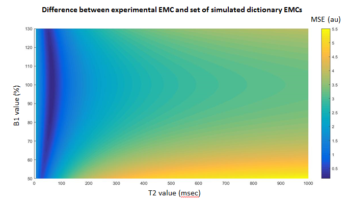
MRI scans: MRI data was acquired on a Siemens 3 Tesla Prisma scanner, for brain anatomy, using MESE protocol and the following scan parameters: NEchoes=20; TE/TR=10/2500 ms; slice thickness=3 mm; in-plane resolution=1.6x1.6 mm2.Gradient-Descent Post-Processing: An EMC dictionary contains a simulated signal curve for each pair of T2 and B1+ values, i.e., EMC (T2, B1+,t). Examining the mean-squared-error cost function for the difference between experimental EMC and each simulated EMC in the dictionary, results in smooth surface, with single global minima (Figure 1). Using this property we implemented a moving-window search scheme across the dictionary two-dimensional search space. Window size of 10x10 was used.
PCA Post-Processing: To compress the data PCA transformation5 was applied on both the simulated EMC dictionary, and on the MESE time course images using the same eigenvectors. Following the transformation, we cropped the number of principle components below 0.1% of the maximal PC, thereby reducing the size of each EMC from the echo train length (ETL) to ~5-6 PCs. This reduced the length of experimental and simulated vectors being compared by a factor of ~3. The cropping threshold was chosen so as to keep the mean relative error and the standard deviation of T2 values below 1 ms.
Analysis: Mean and standard deviation of T2 values were calculated for regions of interest, and relative errors were calculated with respect to T2 values from unaccelerated reconstruction.
Results
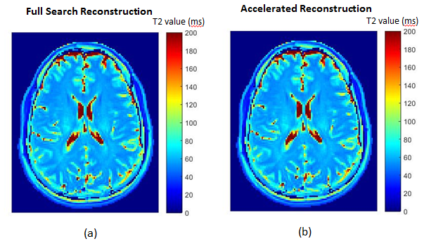
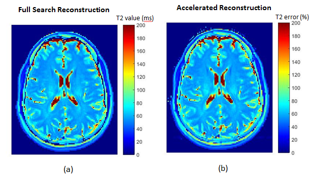
Gradient-decent: Using this method, reconstruction time was reduced approximately x20, with relative errors of < 1% for the brain anatomy. Figure 2 show the resulting T2 maps - the unaccelerated and the accelerated T2 maps.PCA Compression: Figures 3 shows the T2 maps, fitted after PCA compression of the data and search dictionary. Mean ± SD of relative error were 0.67% ± 1.14% for the brain anatomy. Acceleration was much less effective compared to the gradient-descent method, with an average of x1.5 reduction in processing time.
Discussion
Our results demonstrate that acceleration of up to x20 fold can be gained with negligible reduction in accuracy and precision of T2 maps. The may provide suitable means for real-time reconstruction of acquired data. The PCA approach performed less good as compared to the gradient-descent approach, while their combination did not produce significant improvement (not shown). Results are shown for a single slice, and can be expected to scale with the number of slices.Acknowledgements
ISF Grant 2009/17References
1. Eitel I, Friedrich MG. T2-weighted cardiovascular magnetic resonance in acute cardiac disease. J Cardiovasc Magn Reson. 2011;13(1):1-11.
2. Siemonsen S, Mouridsen K, Holst B, et al. Quantitative T2 values predict time from symptom onset in acute stroke patients. Stroke. 2009;40(5):1612-1616.
3. Ben-Eliezer N, Sodickson DK, Block KT. Rapid and accurate T2 mapping from multi-spin-echo data using bloch-simulation-based reconstruction. Magn Reson Med. 2015;73(2):809-817.
4. Shepherd TM, Kirov II, Charlson E, et al. New rapid, accurate T2 quantification detects pathology in normal-appearing brain regions of relapsing-remitting MS patients. NeuroImage Clin. 2017;14:363-370.
5. Pearson K. LIII. On lines and planes of closest fit to systems of points in space . London, Edinburgh, Dublin Philos Mag J Sci. 1901;2(11):559-572.
Figures