3083
Timely Visualizing the Collaterals Formed during Acute Ischemic Stroke with Fe3O4 Nanoparticle-based MR Imaging Probe1Chinese PLA General Hospital, Beijing, China
Synopsis
An αvβ3-specific Fe3O4-RGD nanoprobe was prepared for MR imaging of the collaterals during AIC. The MCAO rat models were constructed for imaging studies on 7.0-T MRI. To show the binding specificity to the collaterals, the imaging results acquired with the Fe3O4-RGD nanoprobe, Fe3O4 mother nanoparticles and RGD blocking experiment. Cerebral ischemia-reperfusion studies were also performed to show the collateral dynamics upon reperfusion. The studies have the first-time enabled the direct observation of collaterals in a quasi-real time fashion and further disclosed that the antegrade flow upon reperfusion dominates the blood supply of primary ischemic tissue during early stage of infarction.
Purpose
Ischemic stroke is one of major leading causes for long-term disability and mortality(1,2). Collateral vessels provide alternative pathway to protect the brain against ischemic injury after arterial occlusion. Aiming at visualizing the collateral vessels occurred during acute ischemic stroke (AIS), an αvβ3-specific Fe3O4-RGD nanoprobe was prepared for in vivo magnetic resonance (MR) imaging of the cerebral collaterals, by taking the advantages of the Fe3O4 nanoparticles in blood vessel MR imaging.Methods
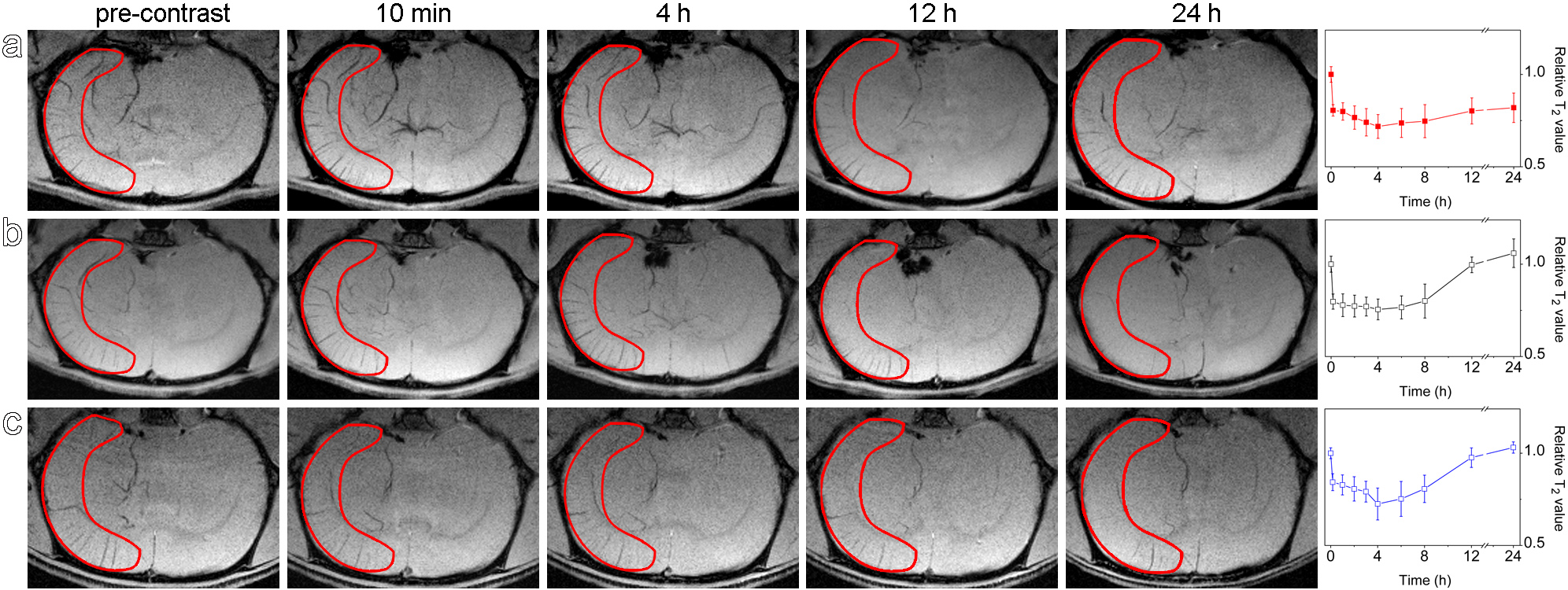
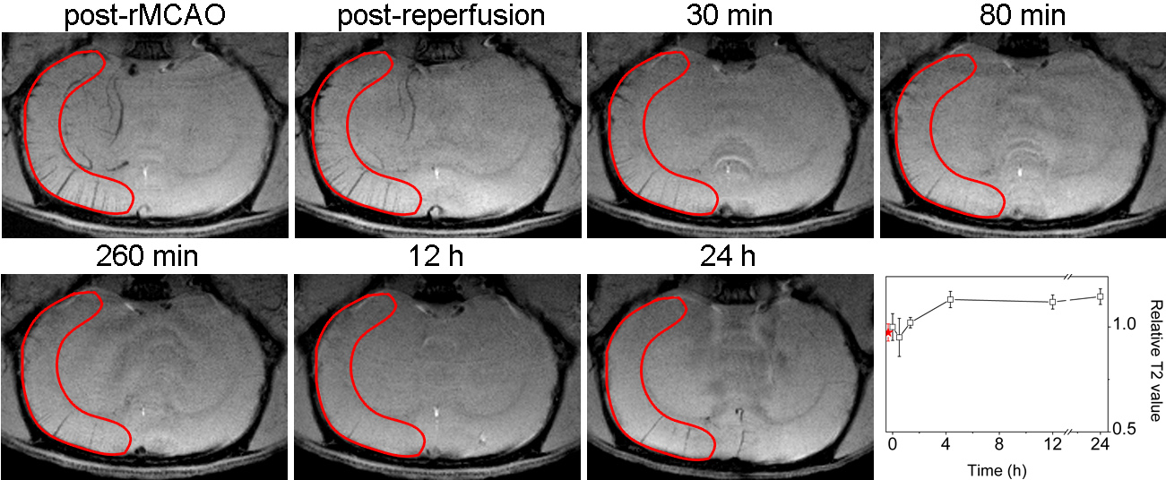
Seventeen adult Sprague-dawley rats of 270 ± 20 g treated for the middle cerebral artery occlusion (MCAO) models.The rats were anesthetized and underwent SWI sequence on 7.0 T MRI systems at serial time points soon after MCAO.The MR imaging experiments on cerebral collaterals were divided into 3groups. The first group of rats received intravenous injection of Fe3O4-RGD nanoprobe shortly after the construction of the rMCAO, the second group received injection of PEGylated Fe3O4 nanoparticles, while the third group received intravenous injection of RGD 30 min prior to Fe3O4-RGD nanoprobe. Following above experiments, cerebral ischemia-reperfusion studies were also performed to show the collateral dynamics upon reperfusion, which is very important for the prognosis of various revascularization therapies in the clinic. Descriptive statistical analysis of the results was conducted for all data analysis.Results
To show the binding specificity to the cerebral collaterals, the imaging results acquired with the Fe3O4-RGD nanoprobes and the Fe3O4mother nanoparticles, respectively, were carefully compared. In addition, RGD blocking experiment was also carried out to support the excellent binding specificity of Fe3O4-RGD nanoprobes. Following above experiments, cerebral ischemia-reperfusion studies were also performed to show the collateral dynamics upon reperfusion, it is found that the occurrence of the cerebral collaterals is a very quick process in acute phase of ischemia stroke, while the disappearance of the cerebral collaterals is also very quick upon reperfusion during the early occlusion of the cerebral artery.Conclusion
The current studies have the first time enabled the direct observation of cerebral collaterals in a quasi-real time fashion and further disclosed that the antegrade flow upon reperfusion dominates the blood supply of primary ischemic tissue during early stage of infarction, which is significantly meaningful for clinical treatment of stroke.Acknowledgements
No acknowledgement found.References
1. Madelung CF, Ovesen C, Trampedach C, Christensen A, Havsteen I, Hansen CK, Christensen H. Leptomeningeal collateral status predicts outcome after middle cerebral artery occlusion. Acta Neurol Scand. 2018, 137, 125.
2. Alves HC, Pacheco FT, Rocha AJ. Collateral blood vessels in acute ischemic stroke, a physiological window to predict future outcomes. Arq Neuropsiquiatr. 2016;74(8):662-670.
Figures