3077
Molecular Targeted Magnetic Resonance Imaging and Spectroscopy1East China Normal University, Shanghai, China
Synopsis
In this work, we
report a novel method for obtaining an exact molecular targeted MRI and MRS.
This method uses the nuclear spin singlet state to select the signals from a
specific molecule. Several endogenetic molecules in living organism such as
N-acetylaspartate and dopamine have been imaged and probed as the targeted
molecules in the MRI and MRS experiments, demonstrating the unique molecular
selectivity of the developed method.
SSS is defined as a nuclear spin state that has a relaxation time much longer than the longitudinal relaxation time T1 and thus often called as the long-lived nuclear spin singlet state.1 An intriguing virtue of SSS, which importance has not gotten enough attention, is its instinctive immunity to the gradient field during evolution.2 By exploiting this property, the evolution period of SSS can be used to select the signals from SSS while suppressing all of the other signals by applying a gradient field. Furthermore, the preparation of SSS of a specific molecule, which requires the unique combination of the chemical shift difference and J-coupling, introduces the salient molecule targeting feature to SSS.3 Combination of this molecule targeting feature and the immunity to the gradient during evolution makes the preparation of SSS possess the perfect molecular level selectivity.
Methods – the pulse sequences for molecular targeted NMR/MRI/MRS
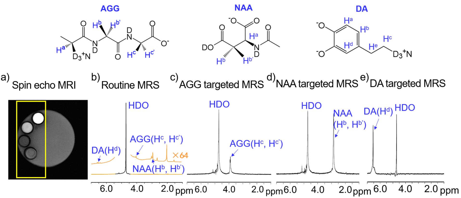
Three pulse sequences designed for the molecular targeted signal selection in nuclear magnetic resonance (NMR), MRI and MRS applications, are exemplarily shown in Figure 1. Figure 1a shows the pulse sequence, YAoWeiXin-NMR, Abbr. YAWX-NMR. The core part of this pulse sequence is the spin singlet state filter, named as SSS filter. Figure 1b shows the scheme of YAWX-MRI pulse sequence, which is designed for molecular targeted MRI. This pulse sequence consists of the SSS filter block to achieve the signal selection of the targeted molecule and the 3D imaging sequence block. Figure 1c shows YAWX-MRS pulse sequence, which is designed for molecular targeted localized MRS. The SSS filter was also introduced into the MRI sequence to achieve a molecular targeted magnetic resonance imaging. To demonstrate this, we designed a special phantom, which was a 5 mm NMR glass tube containing water (40 % D2O and 60% H2O). Four capillaries (1 mm diameter, ~0.1mm wall thickness), containing D2O (60 % D2O and 40% H2O) and the NAA, AGG, and DA aqueous solution, were carefully set into the 5 mm tube.
Results - molecular targeted MRI and MRS
The photo and schematic illustration of this phantom are shown in Figure 2a. Figure 2b shows the routine 1H MRI image of the sample, acquired by using the spin echo imaging sequence. In the image, we can see one big grey disk containing four small disks. Each small disk is surrounded by a black circle. The big grey is from the water inside the 5 mm glass tube, whereas the black, gray and white disks are from the capillaries containing HDO water, the AGG4, NAA and DA aqueous solutions, respectively. The different signal intensity can be attributed to the different 1H concentrations in different solutions. Each small disk has a black ring, showing the wall of the capillary tubes. Using YAWX-MRI, we are able to obtain the molecular targeted images of the sample. Figure 2b, c and d show the molecular targeted 1H MRI image of AGG, NAA and DA respectively, acquired using the pulse sequence in Figure 1b. Figure 3 shows the molecular targeted MRS spectra acquired using the YAWX-MRS sequence. Figure 3a is the MRI image of the sample. The yellow square indicate the region selected for MRS. Figure 3b is the routine 1H MRS spectrum of the selected region in the sample. The signals from the HDO, AGG, NAA and DA can be observed in the spectrum. Figure 3c shows the AGG targeted 1H MRS spectrum. Similarly, the signals of Hb and Hb’ of NAA and Hd of DA are dominant in the NAA targeted and DA targeted 1H MRS spectra of the sample in Figure 3d and 3e, respectively.
Conclusion
In this work we successfully developed three types of pulse sequences. Using these pulse sequences, we have achieved the molecular targeted MRI and MRS, demonstrating the excellent molecular selectivity of the developed methods.
Acknowledgements
This work was supported by National Natural Science Foundation of China (21574043), Microscale Magnetic Resonance Platform of ECNU and the Fundamental Research Funds for the Central Universities.References
1. Carravetta M, Johannessen OG, Levitt MH, Beyond the T1 Limit: Singlet Nuclear Spin States in Low Magnetic Fields, Phys. Rev. Lett.,2004; 92: 153003.
2. Stevanato G, et al., A nuclear singlet lifetime of more than one hour in room-temperature solution. Angew. Chem. Int. Ed., 2015; 54: 3740-3743.
3. Marina C, Levitt MH, Long-lived nuclear spin states in high-field solution NMR. J. Am. Chem. Soc., 2004; 126: 6228-6229.
4. Tayler MCD, Levitt MH, Singlet nuclear magnetic resonance of nearly-equivalent spins. Phy. Chem. Chem. Phy., 2011; 13: 5556-5560.
Figures