2991
A Large FOV 3.0T 16Ch Highly Flexible Array for Body 13C Metabolic Imaging Studies.1GE Healthcare, Inc, Waukesha, WI, United States, 2GE Healthcare, Inc, Aurora, OH, United States, 3University of Wisconsin (Madison), Madison, WI, United States, 4GE Healthcare, Inc, Dallas, TX, United States
Synopsis
Over the last decade, there has been a move toward ultra-flexible conformal coil technology. Similar to flexible H1 coils, there is an interest in developing multi-nuclear coil arrays that are more conformal to human anatomy with highly decoupled elements. In this work, we demonstrate a highly flexible 13C coil array with highly decoupled elements, showing significant improvements in SNR
Purpose
IntroductionThere have been dramatic changes in 1H phased array technology in terms of increasing element density largely driven by the development of parallel imaging methods [1]. At the same time, increasing the density of coil electronics can have negative effects on the coil weight, flexibility and patient acceptance. As such, there has been an interest to achieve light weight and ultra-flexible 1H coils. These coils have been demonstrated [2-3] (called AirTM) using highly decoupled elements. Similarly, multi-nuclear arrays of increasing density are being developing to feed the rise of advanced multi-nuclear imaging methods for 13C, 129Xe and 23Na and other nuclei, and significant progress has been made in the development of the necessary hardware [5-7]. Such methods would greatly benefit from the availability of a large FOV high density multi-nuclear arrays to facilitate multi-organ evaluations. Accordingly, in this work we explored the use of flexible structures to multi-nuclear 13C technology and compare it against existing more rigid conventional designs.
Methods
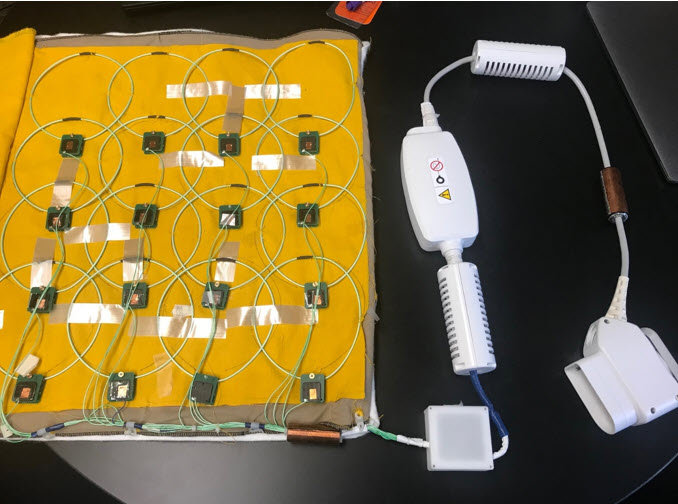
A 16-element blanket 13C array was constructed with outer dimensions 48cm (RL) X 46cm (SI) using a flexible conductor construction of 2.3mm diameter (see Figure 1). Element diameter was 14.5cm to fit the element to element overall of 3.4cm. It was compared against a previously constructed rigid 2 x 4 channel design made form 6cm x 11cm. The Q-factor of the legacy coil was 179 versus 176 for the flexible design with measured improvement of 2.2x B1 sensitivity at a depth of 10cm. Decoupling performance of the flexible loops resulted in a change of -43dB using a single 1” loop sensitivity method. Cable traps for both 127.7MHz and 32.1MHz were attached to the coil. Pre-amplification, decoupling and matching circuitry is included in a discrete module connected to the element loop conductor (see figure 1). The 13C blanket was centered inside of a 13C Tx Clamshell (Rapid Biomedical) on a GE 750W scanner and comparison against rigid 8Ch 13C paddle coils previously described (4). A dimethyl-silicone sphere phantom was utilized for 13C imaging evaluation (5). 2D CSI data were acquired using both receive arrays from a 3 cm slice through the phantom with 48 cm FOV, 16 x 16 imaging matrix, 5000 Hz/1024 pts readout, 500ms TR and 22.5 degree flip angle.Results
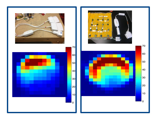
As expected the smaller 6cm x 11cm loops had more B1 sensitivity at the surface of the phantom with the 14.5cm loops having more sensitive at depth. We compared the SNR of the traditional paddle coils with a single row of the 13C array yielding an improvement from SNR of 22 to SNR of 30, giving an overall improvement of 35% (see Figure 2).Discussion
Novel wearable coils are in high interest in both 1H and multi-nuclear imaging. We have demonstrated that it is possible to build large sized highly flexible 13C receive arrays. The coil shows excellent image quality with overall improvement in SNR. This is an important towards future whole body 13C multi-organ imaging.Acknowledgements
The authors would like to thank Josh Kaggie, Martin Graves, Ferdia Gallagher, Mary McLean, Kevin Brindle, Fiona Gilbert, and Evis Sala from the University of Cambridge for their input.References
1. Vasanawala SS, Stormont R, Lindsay S, Grafendorfer T, Cheng JY, Pauly JM, Michael L, Scott G, Guzman JX, Taracila V, Chirayath D, Robb F. Development and Clinical implementation of very light weight and highly flexible Air technology arrays. Proc Intl Soc Mag Reson Med 25 P 0755 2017.
2. McGee KP, Stormont RS, Lindsay SA, Taracila V, Savitskij D, Robb F, Witte RJ, Kaufmann TJ, Huston J, 3rd, Riederer SJ, Borisch EA, Rossman PJ. Characterization and evaluation of a flexible MRI receive coil array for radiation therapy MR treatment planning using highly decoupled RF circuits. Phys Med Biol 2018;63(8):08nt02.
3. “A high-impedance detector-array glove for magnetic resonance imaging of the hand”: Nature Bio Eng., Vol 2570-577 (2018). Zhang B. et al 3."Metabolic Imaging of Patients with Prostate Cancer Using Hyperpolarized [1-13C] Pyruvate" by S. Nelson et al : Sci Transl Med. 2013 Aug 14; 5(198)
4.”Multi-channel metabolic imaging, with SENSE reconstruction, of hyperpolarized [1-13C] pyruvate in a live rat at 3.0 tesla on a clinical MR scanner” by Tropp, J. et al.: Journal of Magnetic Resonance, Volume 208, Issue 1, January 2011, Pages 171-177.
5.” RF instrumentation for same‐breath triple nuclear lung MR imaging of 1H and hyperpolarized 3He and 129Xe at 1.5” by Rao M et al.: Magn Reson Med. 2016 Apr; 75(4): 1841–1848.
6.” Protocol for multi-site quantitative evaluation of 13C radio frequency coils” by Reed G. et al : Abstract # 2510 ISMRM Montreal 2019
Figures