2973
Bloch-Siegert flip angle calibration for phosphorus at the human brain at 9.4 T using ISIS localization1Max Planck Institute for Biological Cybernetics, Tuebingen, Germany, 2Advanced Imaging Research Center, UT Southwestern Medical Center, Dallas, TX, United States
Synopsis
Correct calibration of the transmit field B1+ is crucial to achieve optimal SNR. However, fast and robust B1+ calibration is difficult for X-nuclei due to the low signal sensitivity. In this work, we proposed a fast B1+ calibration method based on the Bloch-Siegert shift and single voxel ISIS localization. With the proposed sequence, the B1+ calibration can be done in less than 5 min for the human brain at B0 = 9.4 T.
Purpose
Fast in vivo flip angle calibration for 31P Magnetic Resonance Spectroscopy in the human brain using Bloch-Siegert pulses with ISIS localization.Introduction
Flip angle calibration is critical to achieve optimal signal-to-noise (SNR) in magnetic resonance spectroscopy. Nevertheless, it is difficult to reliable measure the transmit field strength B1+ for X-nuclei in vivo due to the low sensitivity of the X-nuclei signal. In this work, we present a fast volume selective method to calibrate B1+ and thus flip angles for 31P Magnetic Resonance Spectroscopy (MRS) in the human brain applying a single voxel MRS method called ISIS (Image-Selected in vivo Spectroscopy)1 combined with off-resonant Bloch-Siegert pulses. The Bloch-Siegert shift2 was already used for 31P flip angle calibration at the human heart in combination with a Chemical shift imaging (CSI) sequence3. The advantage of this sequence is the simultaneous acquisition of B1+ values from different locations. However, the needed measurement time was 25 min. The flip angle calibration with the proposed sequence can be done in less then 5 min. For 1H MRS, calibration methods based on single voxel STEAM4 and PRESS5 were presented earlier. However, because of the low T2 values6,7, echo-based methods are not optimal for 31P spectroscopy.The sequence is shown in Fig. 1. The B1+ calculation is based on the phase difference of the PCr resonance introduced by the Bloch-Siegert shift by applying a Fermi pulse with two different frequency offsets3:
$$\gamma B_1 = \sqrt{\frac{(\Phi_2 - \Phi_1) \omega_{RF}}{2 \pi A}}$$
A is the normalized pulse-envelope squared-integral of the Bloch-Siegert pulse, ωRF the frequency offset and φ1,2 the measured phases from the positive and negative frequency offset.
Method
The data was acquired on a 9.4 T whole body MRI scanner (Siemens Healthineers, Erlangen, Germany) with an in-house built double tuned 31P/1H array coil (8TxRx/2Rx for 31P, 10TxRx for 1H)8. All volunteer measurements were done after informed consent.First, the sequence was tested in a phantom measurement by comparing to an established phase sensitive spiral B1+ mapping method9. This method is not suitable for in vivo B1+ calibration because of the extended measurement time needed resulting from the low sensitivity of 31P. The values of the reference B1+ mapping method where averaged over the area of interest covered by the ISIS BS sequence. The following parameters were used for the proposed sequence: voxel size (8 cm)3, TR = 5 s, TE = 0.3 ms, 64 averages, rectangular excitation pulse with TP = 0.35 ms, vector size = 1024, pulse duration GOIA = 5 ms, voltage BS pulse = 250 V, duration BS pulse = 5 ms, frequency offset = ±2500 Hz. The measurement of the ISIS BS was repeated ten times to estimate the variance of the measurement and probe the stability in a phantom.
In a second step, the ISIS BS sequence was tested in vivo in the human brain. First, the reproducibility of the B1+ was probed by repeating the measurement for one volunteer ten times. Second, the B1+ measurement was repeated for seven different volunteers. The parameters were chosen as in the phantom measurement except of a reduction of the number of averages to 16 which reduced the total measurement time to 2.6 min (2x80 sec).
The data evaluation was performed in Matlab by using a self-implemented fitting routine based on the AMARES algorithm10.
Results and Discussion
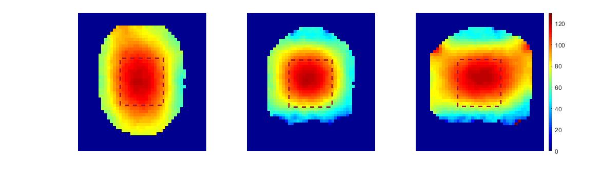
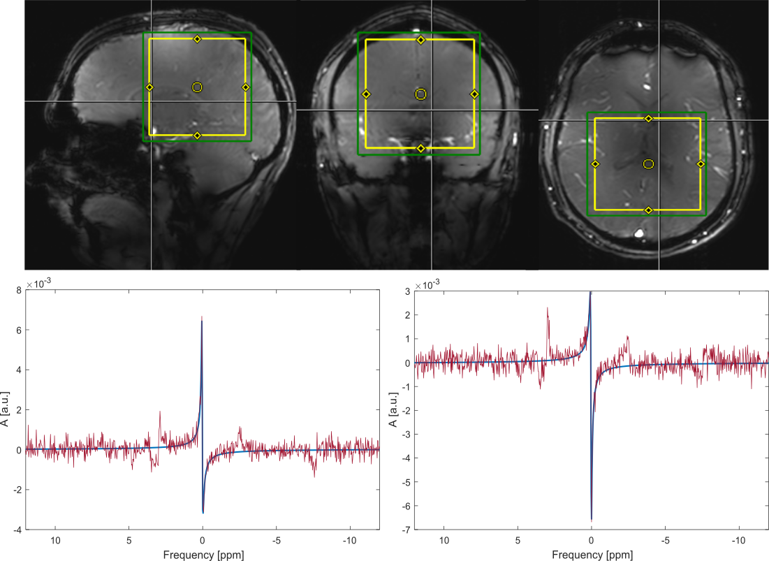
Fig. 2 shows the results of the phase sensitive B1+ sequence. The rectangular area represents the area that was selected with the proposed ISIS BS single voxel B1+ calibration method. For the single voxel sequence, an average B1+ value of (106.43 ± 0.16) nT/V was measured. The corresponding averaged value for the reference method was 101 nT/V. The values only deviate slightly from each other. A reason for that could be that unlike the reference methode the Bloch Siegert sequence measures a weighted average over the area of interest due to different sensitivities of the coil at different positions.Fig. 3 shows the positioning of the voxel in an in vivo measurement and the corresponding spectra for both frequency offsets with the fitted PCr resonance for one volunteer to show spectral quality. No post-processing was applied and the relative phase difference was derived from the two fitted spetra. The reproducibility measurement for ten measurements on a single volunteer resulted in an average measured B1+ of 72.3 nT/V with a standard deviation of 5.3 nT/V.
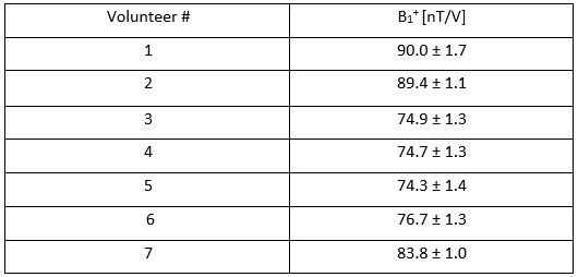
Tab. 1 shows the values for different volunteers. The given standard deviation was calculated from the CRLBs calculated from the fit of the spectra without any post-processing. As known from the reproducibility measurement, this error probably does not reflect the full variation in the measured B1+ values. The higher variance measured in the reproducibility measurement can arise from different reasons as volunteer motion. Also the CRLBs only give a lower estimate of the standard deviation of the fit.
Conclusion
We successfully implemented a single voxel B1+ calibration sequence that can be used for in vivo flip angle calibration for X-nuclei MRS and MRI in less than 5 min. We verified the sequence in phantom measurements and probed a high reproducibility by in vivo experiments in the human brain at 9.4 T for 31P.Acknowledgements
Funding by the European Union (ERC Starting Grant, SYNAPLAST MR, Grant Number: 679927) and the Cancer Prevention and Research Institute of Texas (CPRIT, Grant Number: RR180056) is gratefully acknowledged. Special thanks to Prof. Wolfgang Bogner for his help regarding the ISIS sequence.References
[1] Bogner W, Chmelik M, Andronesi OC, Sorensen AG, Trattnig S, Gruber S. In vivo 31P spectroscopy by fully adiabatic extended image selected in vivo spectroscopy: a comparison between 3 T and 7 T. Magn Reson Med 2011; 66(4): 923 – 930
[2] Sacolick LI, Wiesinger F, Hancu I, Vogell MW. B-1 Mapping by Bloch-Siegert shift. Magn Reson Med 2010; 63: 1315 – 1322
[3] Clark WT, Robson MD, Rodgers CT. Bloch-Siegert B1+-Mapping for Human Cardiac 31P-MRS at 7 Tesla. Magn Reson Med 2016; 76: 1047 – 1058
[4] Versluis MJ, Kan HE, van Buchem MA, Webb AG. Improved Signal to Noise in Proton Spectroscopy of the Human Calf Muscle at 7 T Using Localized B1 Calibration. Magn Reson Med 2010; 63: 207 – 211
[5] Dokumaci AS, Pouymayou B, Kreis R, Boesch C. Motion-Insensitive Determination of B1+ Amplitudes Based on the Bloch-Siegert Shift in Single Voxels of Moving Organs Including the Human Heart. Magn Reson Med 2016; 75: 1867 – 1874
[6] Lei H, Zhu X-H, Zhang X-L, et al. In Vivo 31P Magnetic Resonance Spectroscopy of the Human Brain at 7T: An Initial Experience. Magn Reson Med. 2003; 49: 199 – 205
[7] Van der Kemp JM, Klomp DWJ, Wijnen JP. 31P T2s of Phosphomonoesters, Phosphodiesters, and Inorganic Phosphate in the Human Brain at 7T. Magn Reson Med. 2019; 80 :29 – 35
[8] Avdievich N, Ruhm L, Dorst J, Henning A. Double-Tuned 31P/1H Human Head Array with High Performance at Both Frequencies for Chemical Shift Spectroscopic Imaging (CSI) at 9.4 T. In: Proceed. Of the ISMRM (Montreal) 2019. Abstract nr. 0433.
[9] Allen SP, Morrell GR, Peterson B, Park D, Gold GE, Kaggie D, Bangerter NK. Phase-sensitive sodium B1 mapping. Magn Reson Med 2011; 65(4) : 1125 – 1130
[10] Vanhamme L, van den Boogaart A, Van Huffel S. Improved method for accurate and efficient quantification of MRS data with use of prior knowledge. J Magn Reson 1997; 129(1): 35 – 43
Figures