2969
Perfusion system for studying dynamic metabolomics in neonatal rat brain slices exposed to oxygen deprivation using 1H and 31P NMR1Department of Circulation and Medical Imaging, Norwegian University of Science and Technology (NTNU), Trondheim, Norway
Synopsis
We designed an NMR-compatible bioreactor that allows real-time metabolic measurements of viable rat brain slices under normal conditions and under conditions mimicking hypoxic-ischemic (HI) brain injury. Special emphasis was put on providing physiological temperature in the system, a significant factor in the HI model. 1H NMR spectra were acquired to assess changes in brain metabolites. Peaks of lactic acid, NAA, glutamate, aspartate, and creatine were detected. By acquiring a time series of proton spectra, we observed significantly increased lactate levels over time when switching from normoxia to hypoxia while other metabolites remained stable over the whole experiment.
INTRODUCTION
In vivo animal studies of hypoxic-ischemic (HI) brain injury in the neonatal rat are often hampered by high variability in injury severity, low reproducibility, and many possible confounders related to temperature, feeding etc.1 In vitro oxygen-glucose deprivation (OGD) of neonatal rat brain slices have been shown to reproduce the main pathophysiological events that lead to metabolic impairment in neuroinflammatory diseases like neonatal HI brain injury.2 Tissue slices retain much of the in vivo microenvironment, such as the composition of neighboring cells and supporting structure, while avoiding the confounding factors present in in vivo studies.A bioreactor allows control over temperature, nutrients, and oxygen supply in order to mimic physiological and pathophysiological conditions.3 The aim of this study was to design an NMR-compatible bioreactor that allows for dynamic metabolic measurements of neonatal rat brain slices under normal conditions and under conditions mimicking brain HI, providing the opportunity to learn more about the pathophysiology, and possible effects of treatments. Special emphasis was put on creating a system able to maintain, and monitor physiological temperatures, a feature often overlooked in previous bioreactor designs yet a significant factor in neonatal HI brain injury.
METHODS
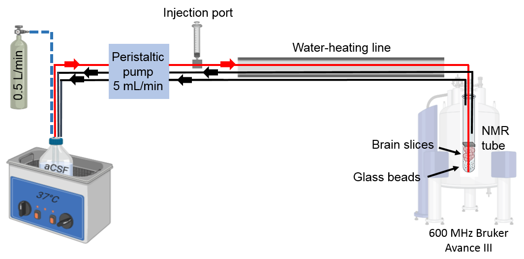
7-day old rat pups were decapitated under anesthesia (3% isoflurane). Brain slices (thickness: 400 µm) were prepared according to the methods in 4. Tissue was loaded into a 10-mm NMR-tube with artificial cerebrospinal fluid (aCSF), and kept in the NMR active region using a sponge filter. Figure 1 shows the design of the bioreactor system. A reservoir of aCSF with 10% D2O (for lock signal) was placed in a water bath outside the NMR spectrometer (temperature set according to experiment). The medium was delivered to the NMR-tube with brain slices using a peristaltic pump (Gilson, Minipuls 3, USA) at a flow rate of 5 mL/min while continuously bubbled with either 95%/5% O2/CO2 gas (normoxia) or 95%/5% N2/CO2 gas (hypoxia) (both from AGA). An active heating using circulating hot water (60 °C) along the supply line was applied.Standard temperature calibrations were performed using a pure MeOH-d4 sample according to 5. In order to calibrate the temperature of the perfused sample inside the magnet and to be able to monitor it during experiments, 0.01% sodium trimethylsilylpropanesulfonate (DSS) was added to the aCSF. The temperature was then determined by measuring the chemical shift distance between water and DSS (DSS set to 0 ppm), and the probe temperature of the NMR spectrometer was corrected to achieve 37 °C in the aCSF sample.
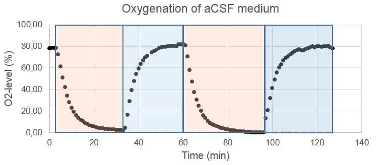
Oxygen levels of the aCSF sample were measured using the ISO-OXY-2 macrosensor (WPI, USA) calibrated according to the manufacturer's instructions. The aCSF reservoir was placed in a water bath set at 37 °C, and bubbled with either 95%/5% O2/CO2 or 95%/5% N2/CO2 gases for around 30 min.
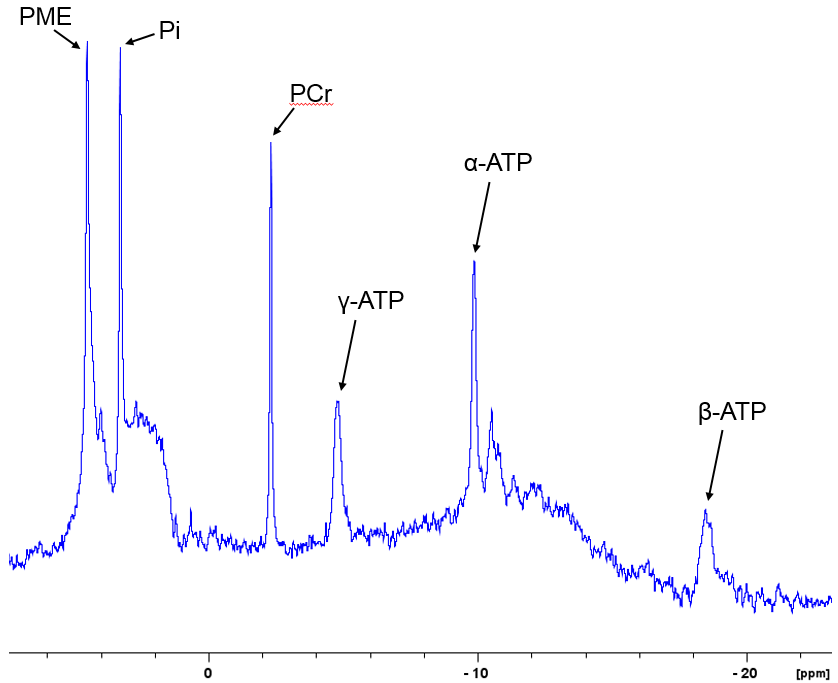
Viability of the neonatal rat brain slices was assessed by 31P NMR (ns = 2400, pulse length = 12.7 μsec). Dynamic changes in metabolites under normoxic and hypoxic conditions were monitored using 1H NMR. A time series of spectra were acquired every 2 min (ns = 32, pulse length = 12.7 μsec). The spectra were phase- and baseline-corrected; lactate peaks were integrated (MatLab, MathWorks), and signal intensity was plotted again time.
RESULTS and DISCUSSION
Recorded oxygen levels in the NMR-tube had a consistently delayed response, taking about 25 min to stabilize at minimum (2%) and maximum (82%) levels after switching between 95%/5% N2/CO2 to 95%/5% O2/CO2 gas supplied to the aCSF (Fig. 2).The initial design of the bioreactor included a temperature of a water bath with aCSF medium, and the NMR spectrometer temperature both set at 37 °C, but lacked any active heating along the supply line. However, the measured temperature of the sample in the NMR-tube with this design was only 24 °C, indicating a heat loss from the tubing. After applying active heating by water circulation along the supply line and increasing the spectrometer temperature, we managed to achieve a stable temperature in the NMR-tube at 37 °C during perfusion.
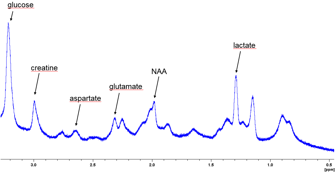
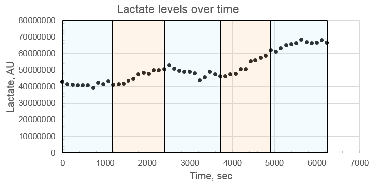
31P NMR was acquired before the oxygen deprivation experiment (Fig. 3). 1H NMR spectra were acquired to assess changes in brain metabolites under normoxia and hypoxia. The peaks of lactic acid, NAA, glutamate, aspartate, and creatine were detected and quantified (Fig. 4). By acquiring a time series of proton spectra, we could follow and quantify the metabolite concentrations over time during the whole experiment with alternating oxygenation levels. We observed significantly increased lactate levels after switching from normoxia to hypoxia in the aCSF (Fig. 5). This is in line with a normal brain tissue response, reflecting increased anaerobic metabolism in the tissue due to hypoxia. Other metabolites remained stable over the whole experiment, probably reflecting the neonatal brain’s resilience against hypoxia alone, and an ability to maintain cell energy production during hypoxic conditions.
CONCLUSION
We have designed an NMR-compatible bioreactor that allows real-time metabolic measurements and maintains the viability of rat brain slices for more than five hours under controlled temperature, oxygenation of nutrients supply. Further work remains to optimize the temporal and spectral resolution of the 1H and 31P NMR protocols in order to monitor a wider range of relevant metabolites.Acknowledgements
The Norwegian University of Science and Technology, Faculty of Medicine and Health Sciences financially supported this work (AM: PhD stipend). MR and animal studies were performed at the MR Core Facility and Comparative Medicine Core Facility at the Norwegian University of Science and Technology.References
1. Rice JE, Vannucci RC, Brierley JB. The influence of immaturity on hypoxic-ischemic brain damage in the rat. Ann Neurol. 1981;9(2):131-141. doi:10.1002/ana.410090206
2. Fernández-López D, Martínez-Orgado J, Casanova I, et al. Immature rat brain slices exposed to oxygen–glucose deprivation as an in vitro model of neonatal hypoxic–ischemic encephalopathy. J Neurosci Methods. 2005;145(1-2):205-212. doi:10.1016/J.JNEUMETH.2005.01.005
3. Gmati D, Chen J, Jolicoeur M. Development of a small-scale bioreactor: Application to in vivo NMR measurement. Biotechnol Bioeng. 2005;89(2):138-147. doi:10.1002/bit.20293
4. Opitz-Araya X, Barria A. Organotypic hippocampal slice cultures. J Vis Exp. 2011;(48). doi:10.3791/2462
5. Findeisen M, Brand T, Berger S. A1H-NMR thermometer suitable for cryoprobes. Magn Reson Chem. 2007;45(2):175-178. doi:10.1002/mrc.1941
Figures