2960
Metabolic profiles of blood plasma in prostate cancer patients having different dietary patterns
Pradeep Kumar1, Rajeev Kumar 2, Sanjay Sharma3, Sanjay Thulkar4, S. Datta Gupta5, S. senthil Kumaran1, Rama Jayasundar1, N. R. Jagannathan6, and Virendra Kumar1
1Department of NMR, All India Institute of Medical Sciences, New Delhi, India, Ansari nagar, India, 2Department of Urology, All India Institute of Medical Sciences, New Delhi, India, Ansari nagar, India, 3Department of Radio-diagnosis, RPC, All India Institute of Medical Sciences, New Delhi, India, Ansari nagar, India, 4Department of Radio-diagnosis, IRCH, All India Institute of Medical Sciences, New Delhi, India, Ansari nagar, India, 5Department of Pathology, All India Institute of Medical Sciences, New Delhi, India, Ansari nagar, India, 6Present address: Department of Radiology, Chettinad Academy of Research and Education, Kelambakkam, Tamil Nadu, India
1Department of NMR, All India Institute of Medical Sciences, New Delhi, India, Ansari nagar, India, 2Department of Urology, All India Institute of Medical Sciences, New Delhi, India, Ansari nagar, India, 3Department of Radio-diagnosis, RPC, All India Institute of Medical Sciences, New Delhi, India, Ansari nagar, India, 4Department of Radio-diagnosis, IRCH, All India Institute of Medical Sciences, New Delhi, India, Ansari nagar, India, 5Department of Pathology, All India Institute of Medical Sciences, New Delhi, India, Ansari nagar, India, 6Present address: Department of Radiology, Chettinad Academy of Research and Education, Kelambakkam, Tamil Nadu, India
Synopsis
In the present study we reported the effect of dietary lifestyle on metabolic profile of blood plasma in prostate cancer (PCa) patients using NMR spectroscopy the long term effects of dietary patterns our metabolic profiles and PCa have been reported in literatures. A significantly higher concentration of choline, glutamate, creatine, succinate, 1-methylhistidine and methionine were observed in non-vegetarians patients compared to vegetarians. Our results suggested that the metabolic pathway alterations seen in amino acids, phospholipids and energy may be related to changes due diet to and PCa progression.
INTRODUCTION
Prostate cancer (PCa) is most frequently diagnosed malignancy in men over the age of 50 years worldwide. It is a slow growing cancer with absence of symptoms at early stages. The pathogenesis of PCa has not been completely understood including the major risk factors associated to cancer development like age, race and family history. Also life style, diet and environmental factors can induce genetic modifications and initiate tumorigenesis processes. Diet is one of the key environmental factors affecting the incidence of various chronic health disorders. Nonveg diet intake has been associated with an elevated risk of several chronic diseases, including diabetes, heart disease and cancers of the colorectal, stomach, esophagus, prostate, breast, and pancreas as well as all-cause mortality1-3. Thus, the present study was carried out to investigate differences in blood plasma of PCa patients and identify biomarker/s associated to the dietary habits of vegetarians and non-vegetarians.METHODS
Blood samples were collected from PCa patients with non-vegetarian (n=43) and from vegetarian (n=25) in morning pre-prandial after overnight fasting. Clinical characteristics of the two groups are presented in Table 1. An informed consent was taken and the study was approved by Institute Ethics Committee. Proton spectra of blood plasma samples were acquired at 700 MHz spectrometer (Agilent, USA) using 1D CPMG with pre-saturation. The following parameters were used for 1D NMR: 64 scan, with a 70s relaxation delay and a spectral width of 9124.1 Hz with an echo time of 15ms. Two dimensional (2D) TOCSY experiment was carried out for assignments of metabolite peaks. For comparison between these two patient groups, unpaired Mann-Whitney U test was carried out using SPSS software. A p-value <0.05 was considered significant. Univariate (receiver operating characteristics (ROC) curve analysis) and multivariate (orthogonal partial least squares–discriminant analyses (OPLS-DA), variable importance to projection (VIP) score statistical analysis was carried out using MetaboAnalyst 4.0.RESULTS
Figure 1 shows the representative aliphatic region of 1D 1H NMR spectrum of blood plasma sample of a PCa patient with non-vegetarian (A) and a vegetarian (B). In all, 29 metabolites were assigned using 1D and 2D NMR. A metabolomics analysis of blood plasma showed an increase concentration of 5 metabolites, glutamate (Glu), creatine (Cr), choline (Cho), succinate (Suc), 1-methyl histidine(1-MH) and lower concentration of methionine (Met) in non-vegetarian PCa patients with as compared to vegetarian. For the concentration [μM (Mean (SD)] of 6 metabolites that showed significant (p<0.05) differences, the AUC values and VIP scores (>1) between the two groups were determined and presented in Table 2. Figure 2 shows the OPLS -DA score plot showing the discrimination of non-vegetarian PCa patients from vegetarian.DISCUSSION
Our data revealed a significantly higher concentration of Cho in the blood plasma of non-vegetarian PCa patients compared to vegetarian. Foods such as meat, meat products, egg yolks and high-fat dairy products are high in phosphatidylcholine and Cho4. Glucogenic amino acid such as Glu was elevated in PCa patients with non-vegetarian diet. Cr is a muscle breakdown product and previous studies have reported higher Cr levels in individuals who consume a non-vegetarian diet (meat)5. Our result also showed significantly higher concentration of 1-methylhistidine with increasing non-veg diets (mainly red meat consumption)4. In contrast, fish eaters had the highest concentrations of amino acids (such as leucine and methionine)6. The ROC curve analysis results indicated that in blood plasma, 3 metabolites with high AUC of above 0.650 (Cr, Met and Suc) while 3 metabolites with less than 0.650 AUC values were seen (such as Glu, Cho and 1-HM). Those metabolites with VIP score (>1.0) were considered potential biomarker/s to discriminate between non-vegetarian patients and vegetarian. MetaboAnalyst 4.0 was used to identify the metabolic pathways which could have been altered in non-vegetarians versus vegetarians. The seven unique significantly altered metabolic pathways seen were arginine and proline metabolism, glycerophosphocholine metabolism, glycine, serine and threonine metabolism, citrate cycle, histidine metabolism, glutamate and glutamine metabolism, and cysteine and methionine metabolism.CONCLUSION
This preliminary study demonstrated that NMR based metabolomics may be useful for distinguishing non-vegetarian PCa patients and vegetarian dietary lifestyle. Further our results provided a better understanding of the metabolic pathway alterations through changes in amino acids, phospholipids, and energy may be associated with diets and PCa progression.Acknowledgements
NRJ thanks SERB, Government of India funding under J.C. Bose Fellowship. PK acknowledges UGC fellowship.References
- Aune D, Ursin G, Veierød MB. Diabetologia. 2009; 52(11):2277-87.
- Micha R, Wallace SK, Mozaffarian D. Circulation. 2010;121(21):2271-83.
- Sinha R, Cross AJ, Graubard BI et al. Arch Intern Med. 2009; 23:169(6):562-71.
- Guasch-Ferré M, Bhupathiraju SN, Hu FB. Clin Chem. 2018;64(1):82-98.
- Goran Gajskia, MarkoGerića, MarijanaVučić et al. Journal of Functional Foods. 2018;48: 643-653.
- Schmidt JA, Rinaldi S, Ferrari P et al. Am J Clin Nutr. 2015;102:1518–26.
Figures
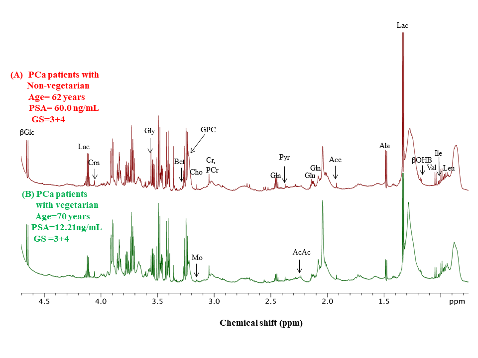
Representative one
dimensional (1D) aliphatic region of 1H-NMR (700 MHz) spectrum of
blood plasma of a non-vegetarian PCa
patient (A) and a vegetarian patient (B).
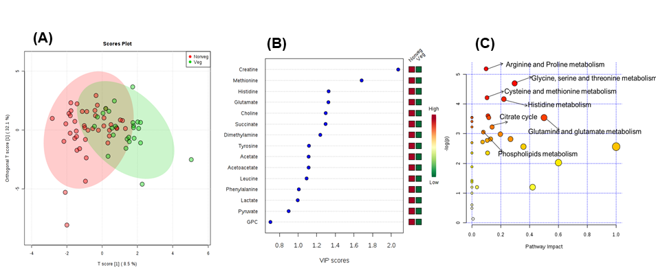
OPLS-DA scores plot
based on blood plasma for the PCa patients with (A) non-vegetarians vs vegetarians,
(B) VIP core plot and (C) overview of metabolic pathway.
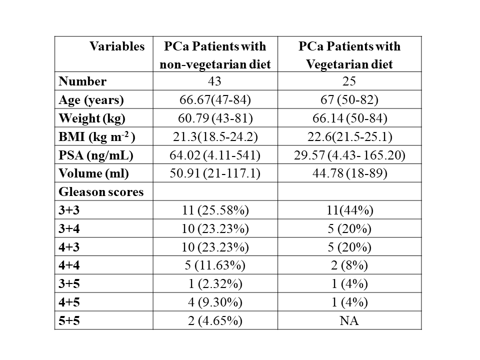
Demographic
and clinical characteristics of the PCa patient with non-vegetarians compared
to vegetarians.
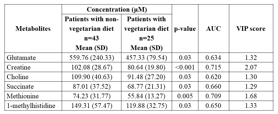
Comparison of the
concentration (μM) of significant metabolites in blood plasma sample of PCa patient non-vegetarians
and vegetarians diets. AUC values were obtained from univariate ROC curve analyses and VIP
score.