2949
MR Spectroscopic Differences of Low and High Grade TERTp-only Gliomas1Institute of Biomedical Engineering, Bogazici University, Istanbul, Turkey, 2Department of Radiology, University Medical Center Utrecht, Utrecht, Netherlands, 3Department of Medical Pathology, Acibadem Mehmet Ali Aydinlar University, Istanbul, Turkey, 4Department of Molecular Biology and Genetics, Acibadem Mehmet Ali Aydinlar University, Istanbul, Turkey, 5Center for Neuroradiological Applications and Reseach, Acibadem Mehmet Ali Aydinlar University, Istanbul, Turkey, 6Department of Neurosurgery, Acibadem Mehmet Ali Aydinlar University, Istanbul, Turkey, 7Department of Radiology, Acıbadem Mehmet Ali Aydinlar University, Istanbul, Turkey
Synopsis
Gliomas that only have the telomerase reverse transcriptase promoter mutation (TERTp-only) are known to have the worst overall survival. The aim of this study was to analyze magnetic resonance spectroscopic (MRS) differences of different grades of TERTp-only gliomas, and classify these groups using machine learning. The results indicated that the ratios of glycerophosphocholine (GPC), glutathione (GSH), total choline (tCho), and glutamine and glutamate complex (Glx) to total creatine increased along with the tumor grade, and machine learning models classified low- and high-grade TERTp-only gliomas with a high accuracy.
Introduction
The importance of genetic biomarkers in the diagnosis and treatment of brain tumors has been emphasized in the 2016 revision of the World Health Organization (WHO) classification of central nervous system tumors1. Isocitrate dehydrogenase (IDH) and telomerase reverse transcriptase promoter (TERTp) mutations, and 1p/19q codeletion have been indicated as genetic biomarkers that affect gliomas’ survival2. Although gliomas that are IDH wild-type but have TERTp mutation (TERTp-only) have been reported to have the worst overall survival2, there have not been any studies investigating the metabolic differences of varying tumor grades within TERTp-only gliomas. The aim of this study is to investigate the MR spectroscopic (MRS) biomarkers of different pathological grades of TERTp-only gliomas, and classify low and high-grade TERTp-only gliomas using machine-learning algorithms.Methods
Fourty glioma patients who have only TERTp mutation (27M/13F, mean age: 59.90±10.44 years, range: 39-84 years, 11 lower grade glioma (grades II and III, LGG), 29 glioblastoma (GBM)) were included in this study. The patients were scanned before surgery at a 3T Siemens Prisma scanner (Erlangen, Germany) using a 32-channel head coil. The brain tumor protocol included pre- and post-contrast (gadolinium DTPA) T1-weighted TSE (TR=500 ms, TE=10 ms), post-contrast T1-weighted volumetric TurboFLASH (TR=2000 ms, TE=4 ms), post-contrast T2-weighted TSE (TR=5000 ms, TE=105 ms), and T2*-weighted gradient-echo echo planar imaging (EPI) dynamic susceptibility contrast (DSC) MRI (TR=1500 ms, TE=30 ms). MRS data were acquired from the solid tumor region excluding necrosis, edema, and hemorrhage using a Point Resolved Spectroscopy (PRESS) sequence (TR/TE=2000/30 ms, voxel size=1-8 cm3). LCModel spectral fitting program3 was used for quantification of MR spectroscopic peak concentrations of 19 metabolites including glycerophosphocholine (GPC), glutamate (Glu), glutamine (Gln), and glutathione (GSH), and five composite peak concentrations [total choline (tCho=GPC+PCh), total creatine (tCr=Cr+PCr), total N-acetyl aspartate (tNAA=NAA+NAAG), glutamine and glutamate complex (Glx), and myoinositol and glycine (mIns+Glyc)]. The metabolites, which have a Cramer-Rao lower bound (CRLB) of more than %30, were excluded from the study. Afterwards, the metabolite intensities were normalized to tCr. A Mann-Whitney U test was used to assess metabolic differences between low- and high-grade TERTp-only gliomas. Bonferroni multiple comparison correction was applied, and a P value of less than 0.002 was considered as statistically significant. Afterwards, machine-learning algorithms were used to classify low- and high-grade TERTp-only gliomas based on their metabolic profiles in Classification Learner app of MATLAB 2019a (The MathWorks Inc., Natick, MA). The classification was first conducted using all metabolites, and then using only the metabolites which differed between the two groups. In both classification tasks, 5-fold cross validation was used, and the models were executed 100 times to report the mean performance measures.Results
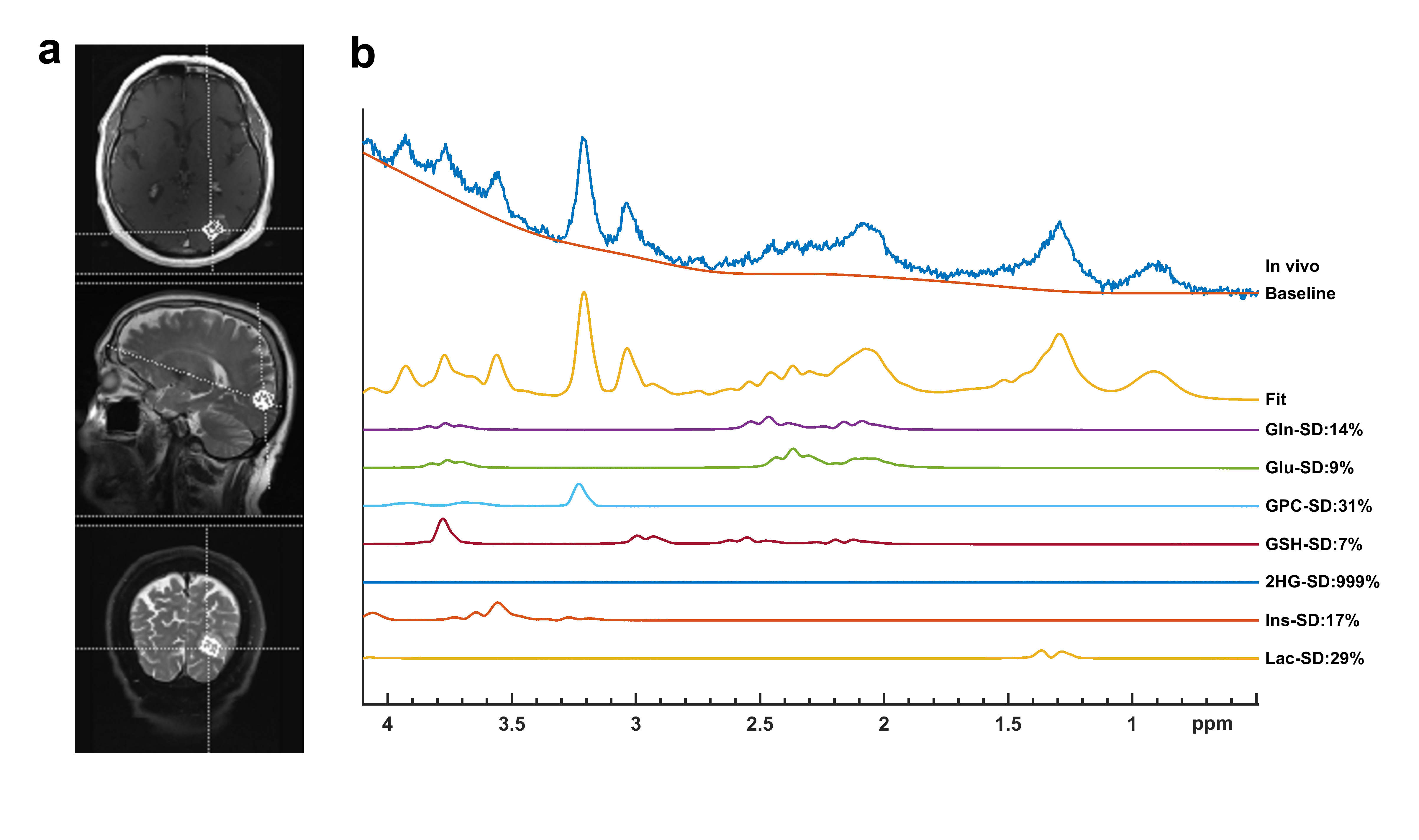
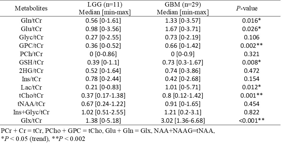
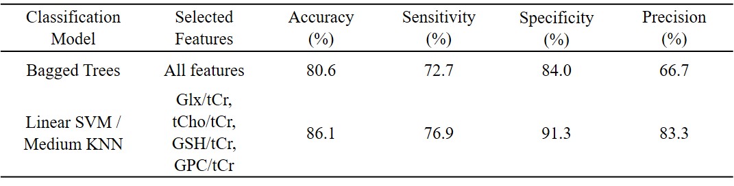
Figure 1 shows an example TERT-only GBM patient’s short-TE PRESS MRS data along with (a) voxel selection on T2-weighted TSE and (b) some of the LCModel quantification results. The median and range of normalized metabolite concentrations and the P-values of their comparison between low- and high-grade TERTp-only gliomas are shown in Table 1. TERTp-only GBM patients had a statistically significantly higher GPC/tCr (P=0.002), tCho/tCr (P=0.001), and Glx/tCr (P<0.001) than TERTp-only patients with lower grades. There was also a trend for a higher GSH/tCr (P=0.008) in GBM than LGG. The classification results of the models giving highest accuracy are summarized in Table 2. A bagged trees model was the best one for the classification using all features resulting in an 80.6% accuracy, 72.7% sensitivity, 84% specificity, and 66.7% precision. The classification performance was improved by feature selection, and the best performance results were obtained using either a linear SVM or a medium KNN model with Glx/tCr, tCho/tCr, GSH/tCr, and GPC/tCr, resulting in an accuracy of 86.1%, sensitivity of 76.9%, specificity of 91.3%, and precision of 83.3%.Discussion and Conclusion
The results of this study indicate that GPC/tCr, GSH/tCr, tCho/tCr and Glx/tCr ratios increase in high-grade TERTp-only gliomas. These metabolites have recently been indicated as important biomarkers of TERTp-only gliomas4. Moreover, machine learning models were successful in differentiating the pathological grades using these metabolic markers. Since overall survival is worse in high-grade gliomas, the metabolic profile of TERTp-only GBM implies that high glutamate and choline species, and GSH might be indicative of poor prognosis. These results could be improved by using a larger patient cohort and by optimizing the parameters of machine learning models.Acknowledgements
This study has been supported by TUBITAK 1003 grant 216S432.References
1. Hoshide R, Jandial R. 2016 World Health Organization Classification of Central Nervous System Tumors: An Era of Molecular Biology. World Neurosurg. 2016; 94: 561-562.
2. Eckel-Passow JE, Lachance DH, Molinaro AM, et al. Glioma Groups Based on 1p/19q, IDH, and TERT Promoter Mutations in Tumors. The New England journal of medicine. 2015; 372: 2499-2508.
3. Provencher SW. Automatic quantitation of localized in vivo 1H spectra with LCModel. NMR Biomed. 2001; 14: 260-264.
4. Ozturk-Isik E, Cengiz S, Ozcan A, et al. Identification of IDH and TERTp mutation status using (1) H-MRS in 112 hemispheric diffuse gliomas. J Magn Reson Imaging. 2019.
Figures