2938
Effects of lisinopril on arterial stiffness, hippocampal blood flow, N-acetyl aspartate and cortical thickness in hypertensive Dahl-s rats
Samuel Ajamu1, Rachel Fenner1, Nikkita Khattar2, Yulia Grigorova1, Edward Lakatta1, Ondrej Juhasz1, Peter Rapp3, Mustapha Bouhrara2, Richard Spencer2, Olga Fedorova1, and Kenneth Fishbein2
1Laboratory of Cardiovascular Science, National Institute on Aging, Baltimore, MD, United States, 2Laboratory of Clinical Investigation, National Institute on Aging, Baltimore, MD, United States, 3National Institute on Aging, Baltimore, MD, United States
1Laboratory of Cardiovascular Science, National Institute on Aging, Baltimore, MD, United States, 2Laboratory of Clinical Investigation, National Institute on Aging, Baltimore, MD, United States, 3National Institute on Aging, Baltimore, MD, United States
Synopsis
Central arterial stiffness (CAS), associated with hypertension, is likely associated with attendant cerebral hypoperfusion, neuronal density loss and cognitive decline, and stiffening of cerebral arterial wall. We previously found associations between pulse wave velocity (PWV), a marker of CAS, and hippocampal cerebral blood flow (CBF) and neuronal density in 6 months old hypertensive Dahl salt-sensitive (Dahl-S) rats, which exhibit age-associated memory loss. The present study showed the ACE inhibitor, lisinopril, resulted in stabilized hippocampal blood flow and NAA concentration compared to nontreated age-matched animals. We also observed significant changes in cortical thickness for treated animals compared to nontreated control.
Introduction
Hypertension and diseases of the vascular wall may be significant contributors to dementia, including Alzheimer’s disease.1,2,3 Obtaining an effective treatment for vascular dementia remains an elusive goal, with the mechanisms of these processes remaining poorly understood. Central arterial stiffness (CAS), as reflected by increased pulse wave velocity (PWV), is a state in which the compliance of large blood vessels, including the aorta, decreases, secondary to remodeling induced by systemic hypertension. It remains unknown whether this is accompanied by increased stiffness in the cerebral arteries, and if so, whether there are consequences related to prefusion of critical cerebral structures. We have previously shown that CAS variability was strongly correlated to differences in hippocampal blood flow and NAA concentrations in hypertensive Dahl-S rats.4 To further understand the role of CAS in cerebral perfusion and its sequelae, we used an ACE inhibitor to modulate CAS in hypertensive Dahl-S rats.Methods
Male Dahl-S rats (SS/JrHsd; n=32; 14 treated, 18 untreated; Charles River Laboratories) were fed with a normal salt diet (0.5% NaCl) until age 22 weeks, when systolic blood pressure (SBP) and PWV were measured using the procedure described in Ajamu et al.4 Lisinopril (15mg/kg of body weight/day)5 was administered starting six-months post baseline scans. Subsequent measurements were taken after 3 and 6 months of treatment. N-acetyl aspartate (NAA) concentration was measured in the right hippocampus using a 7T Bruker Biospec MRI scanner within a 2.5mm×1.5mm×2.5 mm spectroscopic voxel using a CHESS-PRESS sequence with a TE/TR=15.7 ms/1s. The concentration of NAA, an indicator of neuronal density6, was calculated using LCModel. Cerebral blood flow (CBF) was measured using continuous arterial spin labeling (CASL) in the coronal slice with the largest hippocampal area. Data were acquired with an EPI sequence with parameters TE/TR= 28ms/5s, 128 averages, 0.469mm×0.469mm×1mm voxel size and labeling time of 2 s. CBF was estimated in in the hippocampus after NESMA filtering of images7 according to standard method.7,8,9 Cortical Thickness (CT) analysis was done using a T2 weighted RARE 3D image (n=12). The T2 weighted rare images were acquired with a TE/TR= 38 ms/1s, 0.2mm×0.2mm×0.2mm voxel size for each animal at each timepoint. Advanced Normalization Tools (ANTs) were used for linear and nonlinear registration. Images were rigidly aligned with a pre‐existing atlas (Waxholm Sprague-Dawley atlas) using mutual information for study.The cortical label from the atlas was used to segment out each subject’s cortical region and cortical thickness was performed using ANTs protocols.10,11Results
At 6 months of age, the Dahl-S rats displayed moderate hypertension (SBP: 164±13; as compared to a 3 months baseline of 142±5 mmHg; mean±SD; P<0.01, t-test). SBP of nontreated (NT) animals increased at all subsequent time points while treated animals exhibited a decrease in SBP from 6-9 months of age. SBP was stable low from 9-12 months of age (Figure 1a). In treated animals, PWV decreased at all subsequent timepoints after 6 months. For NT animals, PWV remained steady from 6-9 months but increases from 9-12 months (Figure-1b). Hippocampal CBF of NT animals decreased in a downwards trend but remain stabilized in the treated group across timepoints (Figure-2a). Likewise, the hippocampal NAA, among treated animals remained stabilized, while in NT animals this parameter fluctuated with an eventual downwards trend. The mean difference maps of the cortical thicknesses for the NT and treated rats are similar except for the regions denoted by the red arrows when comparing treated to nontreated.(Figure-3) The occipital region showed cortical thickening (blue) and parietal region showed cortical thinning (yellow). The p value maps show that those regions of significant thickening and thinning are greater in the treated group compared to the NT Dahl-s rats.(Figure-4)Discussion
We found that the decrease in CAS secondary to lisinopril treatment served to maintain levels of hippocampal CBF and neuronal mass, as well as potentiate cortical thickening in the occipital region of Dahl-S rats. While numerous studies have linked hypertension with CAS, relatively little is known about the relationship between CAS and neuronal mass. The present study is a substantial expansion of our previous work, in which we found that increased CAS was associated with lower hippocampal perfusion, indicating a potential pathophysiologic mechanism for impaired neuronal function in hypertension and a possible link with cognitive impairment. Here, we sought to decrease CAS with an ACE inhibitor, lisinopril, which attenuated the age-associated increase in CAS5 and decrease in hippocampal blood flow. In addition, treatment served to prevent the loss of hippocampal NAA that occurred after 9 months of age. Finally, we found occipital cortical thickness exhibited a greater increase in lisinopril-treated animals at older ages. Given that the posterior cerebral artery is a dominant source of perfusion to the hippocampus12 and the occipital lobe13, these findings suggest a coherent pattern through which cerebral arteries decrease in stiffness and improved regional CBF. Therefore, CAS may be a therapeutic target for vascular dementia.Conclusion
In the Dahl-S rat model of age-associated hypertension, lisinopril served to attenuate vascular effects that drive decreased CBF in hypertension. As compared to controls, treatment resulted in greater hippocampal NAA and increased occipital cortical thickness. This motivates further consideration of appropriate treatment of hypertension and vascular wall disease as a therapeutic target for dementia.Acknowledgements
The work was supported by the Intramural Research Program of the National Institute on Aging of the National Institutes of Health.References
- Iadecola C, Davisson RL. Hypertension and cerebrovascular dysfunction. Cell Metab 2008;7(6),476-84.
- Mitchell GF, Effects of central arterial aging on the structure and function of the peripheral vasculature: implications for end-organ damage. J Appl Physiol (1985), 2008; 105(5), 1652-60.
- Mitchell GF, et al. Arterial stiffness, pressure and flow pulsatility and brain structure and function: the Age, Gene/Environment Susceptibility--Reykjavik study. Brain 2011;134(Pt 11), 3398-407.
- Ajamu S, Association of Central Arterial Stiffness with Hippocampal Blood Flow, N-Acetyl Aspartate and Anxiety in Hypertensive Dahl Salt Sensitive Rats. Alzheimer’s & Dementia 2019;15(7), 279-280.
- Brilla CG, Janicki JS, Weber KT. Cardioreparative Effects of Lisinopril in Rats With Genetic Hypertension and Left Ventricular Hypertrophy. Circulation 1991;83:1771-1779.
- Baslow MH. N-acetylaspartate in the vertebrate brain: metabolism and function. Neurochem Res 2003;28:941-53.
- Bouhrara M, Lee DY, Rejimon AC, Bergeron CM, Spencer RG. Spatially adaptive unsupervised multispectral nonlocal filtering for improved cerebral blood flow mapping using arterial spin labeling magnetic resonance imaging. J Neurosci Methods 2018;309:121-131.
- Lu H, Leoni R, Silva AC, Stein EA, Yang Y. High-field continuous arterial spin labeling with long labeling duration: reduced confounds from blood transit time and postlabeling delay. Magn Reson Med 2010;64(6),1557-66.
- Barbier EL, Lawrence KS, Grillon E, Koretsky AP, Décorps M. A model of blood–brain barrier permeability to water: Accounting for blood inflow and longitudinal relaxation effects. Magn Reson Med 2002;47:1100-1109.
- Pagani, M., Damiano, M., Galbusera, A., Tsaftaris, S. A., & Gozzi, A. (2016). Semi-automated registration-based anatomical labelling, voxel based morphometry and cortical thickness mapping of the mouse brain. Journal of neuroscience methods, 267, 62-73.
- Vetreno, R. P., Yaxley, R., Paniagua, B., Johnson, G. A., and Crews, F. T. (2017) Adult rat cortical thickness changes across age and following adolescent intermittent ethanol treatment. Addiction Biology, 22: 712– 723.
- Erdem, A., Yaşargil, M., & Roth, P. (1993). Microsurgical anatomy of the hippocampal arteries, Journal of Neurosurgery, 79(2), 256-265.
- Jong S. Kim, Posterior Cerebral Artery Disease, Stroke,2016,393-412.
Figures
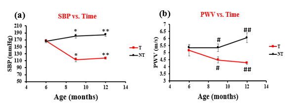
Longitudinal changes in cardiovascular parameters in Dahl-S rats treated with
lisinopril
Systolic
blood pressure (SBP) (a) and pulse wave velocity (PWV) (b), in
lisinopril treated (T) and nontreated (NT) animals. SBP between NT and T animals
at 9 and 12 months were
significantly different ( *p < 0.001 and ** p < .001, respectively). This
shows the efficacy of the drug as a treatment of systolic hypertension. PWV
between NT and T animals at 9 months were significantly different (#p = 0.04).
Similarly, at 12 months there is a significant difference in the groups PWV (##p
< 0.001 ).
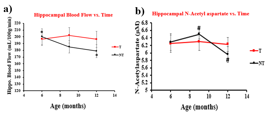
Longitudinal changes in cerebral
parameters in Dahl-S rats treated with lisinopril
Hippocampal
CBF (a) and NAA (b) in lisinopril T and NT Dahl-S rats. Though there is no
significance for hippocampal blood flow in the two groups, a declining trend emerges
in NT rats while T rats stabilized. Values at 6 vs.12 months in the NT group were
borderline significant (*p=0.06, paired t-test). There was also no significance
in NAA between groups. But, NAA for the NT group from 9 - 12 months show a
downward trend, suggesting lower neuronal density compared to the T rats (#p =
0.07,paired t-test).
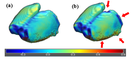
Mean Differences in Cortical Thickness. (Longitudinal
brain MRI measurement; 6 – 12months) [6-month CTs subtracted by 12-month CTs]
Mean difference between age six and twelve
months of (a) nontreated animals and (b) lisinopril treated animals. Blue
indicates regions of thickening while yellow indicated regions of thinning.
Occipital and parietal regions indicated by the red arrows. Despite the PCA
feeding the medial parietal region, many other vessels are responsible for its adequate
perfusion.
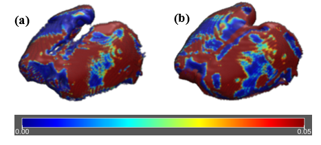
P value map comparing longitudinal changes
in cortex thickness in six and twelve months old nontreated animals (a) and
lisinopril treated animals (b). All insignificant p values (> 0.05) were revalued
as 0.05 for scaling visualization purposes. This reveals that the regions of thickening
found in the occipital cortical region and the thinning found in the temporal
cortical region are significant. Compared to the nontreated animals the
significant regions are relatively larger.