2935
Diurnal fluctuation of glutathione measured with MR spectroscopy1Human Brain Research Center, Kyoto University, Kyoto, Japan, 2Department of Psychiatry, Kyoto University, Kyoto, Japan, 3National Institute for Physiological Sciences, Okazaki, Japan
Synopsis
Glutathione (GSH) is a powerful and general antioxidant found in cell of various animals. Since its concentration in central nervous system has been thought to related to psychiatric disease, several studies has carried out but the results are still controversial. Here, we investigated GSH diurnal fluctuation of healthy subjects and compared with the results of the reproducibility study. Our results shows GSH concentration in brain fluctuates by 10 to 20% during the daytime. This finding indicates that conditions of MRS examination must be regulated strictly and the fluctuation should be took into account at interpretation of GSH studies.
Introduction
Glutathione (GSH) is a powerful and general antioxidant found in cells of animals, plants, and so on. GSH exists in various cells also in the human body and protects from attacks of reactive oxygen species such as free radicals invading into the cell. Therefore, GSH concentration in the central nervous system has been thought to relate tolerance against stress, and so several MR spectroscopy (MRS) studies have been carried out to compare GSH concentration between healthy subjects and psychiatric patients invasively 1,2. However, the results are controversial so far. There are two possible reasons; one is that GSH detection with MRS is basically difficult, i.e., reproducibility is low, and the other is that diurnal fluctuation of GSH may be affected. Considering that diurnal fluctuation of GSH concentration in human blood has already been reported 3,4, that in the human brain can be happened. Therefore, here we investigated GSH fluctuation of healthy subjects during the daytime.Methods and Materials
We have gotten an approval from the local IRB on this study. Two studies were carried out; one is on diurnal fluctuation and the other is for reproducibility evaluation to validate our methodology. For the former study, a totally of eight normal subjects (two 20s’/30s’ and males/females, respectively) were scanned six times in a day with 2.5 hours intervals. They were asked to come without breakfast in the morning and to have meals between 1st and 2nd (breakfast), 3rd and 4th (lunch), and 5th and 6th scans (dinner). For the latter study, ten normal subjects (24 to 44 y.o., male) were scanned two times successively with a 10 to 15 minutes break.A whole-body 3T scanner, Trio Tim (Siemens, Erlangen, Germany) is used with 32ch phased-array head coil. MRS sequences we used including single-voxel PRESS and fastestmap were provided by Minnesota University (TR version). Prior to the PRESS scan, shimming with fastestmap (FWHM < 13 Hz), RF power adjustment, VAPOR (water suppression) parameter adjustments were carried out carefully as instructed in the documents of the sequences. The voxel size was 20x20x30 mm and placed at the Precuneus region. TR and TE were 3000, 30 msec, respectively and Nex was 128.
Measured data were analyzed with LCModel 5,6 and the estimated GSH concentration was normalized with Cr+pCr.
Results and Discussion
Totally sixty-seven PRESS scans (47 scans for the diurnal fluctuation study and 20 scans for the reproducibility study. Only one scan of the former study was skipped due to time limitation) were carried out and, on all of the data except one, values of Cramer-Rao lower bound (CRLB) were less than 15%, i.e., the data quality was sufficient to discuss.Table 1 shows the reproducibility results. Coefficients of variance were up to 7%, and its average was 2.14%.
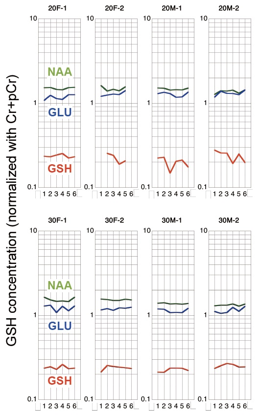
Figure 1 shows the diurnal fluctuation of GSH with that of NAA and glutamate as references. Comparing with the reproducibility of around 2%, the fluctuation during a day of up to 10 to 20% is sufficiently high to indicate the existence of the fluctuation. Its tendency among the subjects is not so obvious, but five of the eight subjects (one 20’s female and four 30s') seem to show a similar change, i.e., GSH is low in the morning and evening, and high in the daytime. Interestingly this is opposite from the tendency of GSH in blood 1,2.
Considering the sufficiently low CRLB of our data and the high reproducibility of our method, our results strongly suggest that diurnal fluctuation is not negligible. Therefore, at the planning of the GSH study, we should carefully consider conditions for the examination, and the obtained data must be interpreted with consideration of this diurnal fluctuation.
Acknowledgements
We express our sincere thanks to Dr. Lana Kaiser and Mr. Katsutoshi Murata (Siemens Healthineers Japan, Japan) for great help to use the sequence. We would like to acknowledge Edward J. Auerbach, Ph.D. and Małgorzata Marjańska, Ph.D. (Center for Magnetic Resonance Research and Department of Radiology, University of Minnesota, USA) for the development of the pulse sequences for the Siemens platform which were provided by the University of Minnesota under a C2P agreement. This research was supported by JSPS KAKENHI Grant No.15K09826References
1. Terpstra, M et al., Validation of glutathione quantitation from STEAM spectra against edited 1H NMR spectroscopy at 4T: application to schizophrenia, MAGMA (2005) 18: 276–2822.
2. Wang, AM, et al., Assessing Brain Metabolism With 7-T Proton Magnetic Resonance Spectroscopy in Patients With First-Episode Psychosis, JAMA Psychiatry. 2019;76(3):314-3233.
3. Valencia E, et al., Circadian rhythmicity of whole-blood glutathione in healthy subjects., Nutrition. 2001 Sep;17(9):731-3.4.
4. Blanco RA1, et al., Diurnal variation in glutathione and cysteine redox states in human plasma., Am J Clin Nutr. 2007 Oct;86(4):1016-23.5.
5. Provencher, SW, Estimation of metabolite concentrations from localized in vivo proton NMR spectra, Magn. Reson. Med., 30 (6) (1993), 672-6796.
6. Provencher, SW, Automatic quantitation of localized in vivo 1H spectra with LCModel, NMR Biomed., 14 (4) (2001), 260-264
Figures
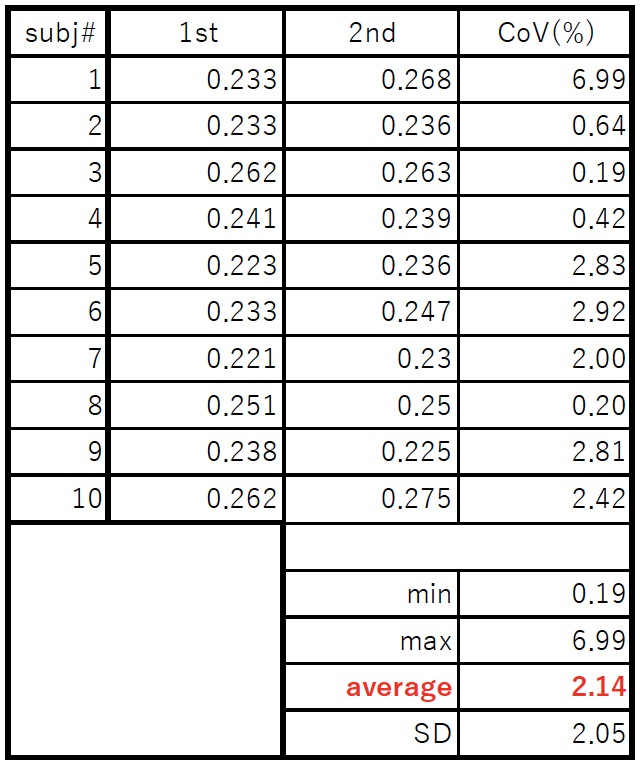
table1
The results of reproducibility results.