2926
A 9-month follow-up study in patients with delayed neurologic sequelae after CO intoxication using absolute quantification of 1H MRS and DKI
Yi-Chen Tsai1, Hui-Ru Tsai1, Ming-Chung Chou2, Ping-Hong Lai3,4, Jie-Yuan Li5,6, and Cheng-Wen Ko1
1Dept. of Computer Science and Engineering, National Sun Yat-sen University, Kaohsiung, Taiwan, 2Dept. of Medical Imaging and Radiological Sciences, Kaohsiung Medical University, Kaohsiung, Taiwan, 3Dept. of Radiology, Veterans General Hospital-Kaohsiung, Kaohsiung, Taiwan, 4School of Medicine, National Yang-Ming University, Taipei, Taiwan, 5Dept. of Neurology, E-Da Hospital, Kaohsiung, Kaohsiung, Taiwan, 6School of Medicine, I-Shou University, Kaohsiung, Kaohsiung, Taiwan
1Dept. of Computer Science and Engineering, National Sun Yat-sen University, Kaohsiung, Taiwan, 2Dept. of Medical Imaging and Radiological Sciences, Kaohsiung Medical University, Kaohsiung, Taiwan, 3Dept. of Radiology, Veterans General Hospital-Kaohsiung, Kaohsiung, Taiwan, 4School of Medicine, National Yang-Ming University, Taipei, Taiwan, 5Dept. of Neurology, E-Da Hospital, Kaohsiung, Kaohsiung, Taiwan, 6School of Medicine, I-Shou University, Kaohsiung, Kaohsiung, Taiwan
Synopsis
In this study, we explored the metabolic changes in WM of patients with and without DNS using 1H MRS and DKI longitudinally at 1 week and 1, 3, and 9 months after CO intoxication. Increased Cr and Cho were observed at early stage in WM of patients with DNS. Decreased tNAA showed higher correlation with the presence of WM lesion than of DNS. The chronic change of Ins and Glx in patients with DNS implies themselves as potential indices to provide valuable information for monitoring DNS development.
Introduction
Delayed neurologic sequelae (DNS) is commonly developed in patients with CO intoxication, which exhibits recurrent neuropsychiatric symptoms after an interval of apparent normality1,2. A neural marker for DNS is thus desirable to facilitate prediction and monitoring of neuropathology for appropriate diagnosis and treatment2,3. In this study, we investigated the metabolic change in WM of patients with DNS at 1 week and 1, 3, and 9 months after CO intoxication using absolute quantification of 1H magnetic resonance spectroscopy (MRS). Voxel-based diffusion kurtosis analysis was included for comparison4.Methods
A total of 24 patients who experienced CO intoxication were enrolled in this study with IRB approval. Eleven patients who exhibited white matter lesion (WML) within follow-up period were categorized as WML+ and others as WML-. In WML+ group, 5 patients were diagnosed with the presence of DNS (DNS+), and others as DNS-. All patients were evaluated regularly at 1 week and 1, 3, 9 months after CO intoxication. 25 normal subjects were included as control group.All MR studies were conducted on a 1.5T system (General Electric) using 8-ch head coil with single voxel spectroscopy protocol (PRESS, TR/TE = 1600/35 ms, Ave =128, voxel size = 2x2x2 cm3). By referencing to axial T2 images, MRS voxel was placed on the left occipital WM region where WM abnormalities of DNS frequently occurred (Fig.1a). Acquired spectra were analyzed by LCModel to compare the absolute metabolic levels of NAA+NAAG (tNAA), Creatine (Cr), Choline (Cho), Myo-Inositol (Ins) and Glutamate+Glutamine (Glx). 3D axial T1 images were used for white matter (WM), gray matter (GM) and cerebrospinal fluid (CSF) segmentation by SPM12. The fractions of WM, GM and CSF within voxel of interest (VOI) were applied to LCModel analysis for partial volume correction. Cramér-Rao lower bounds (SD%) of values larger than 20% were excluded in the statistical analysis. Absolute concentrations were presented as mean and standard deviation in mmol/mL (institutional unit). Whole-brain DKI data were acquired using a twice-refocused spin-echo diffusion-weighted pulse sequence with TR/TE=10000/91ms, b-values=1000 and 2000 s/mm2, number of b0 images=4, matrix size=80x80, FOV=240x240 mm2. Parameters of fractional anisotropy (FA), axial kurtosis (AK), radial kurtosis (RK) and mean kurtosis (MK) were calculated in the corresponding VOI of MRS. Non-parametric Mann–Whitney U test was applied to group analysis where the p-value < 0.05 was considered statistically significant.
Results and Discussion
Quantitative comparison of tNAA, Cr, Cho, Ins and Glx between patients with/without DNS are demonstrated in Fig.1. The tNAA of WML+DNS+ patients are dramatically decreasing from 10.58±3.51 mmpl/mL at 1 week to 6.36±1.03 mmpl/mL at 1 month. Both of Cr and Cho in patients with DNS (WML+DNS+) show significant increase at 1 month (p<0.05) whilst Ins increases significantly at late stage from 3 months to 9 months (p<0.05).DKI indices (FA, AK, MK and RK) derived from VOI of MRS demonstrate that they performed well in differencing patients with DNS from DNS- ones (Fig.2). Patients with DNS had higher diffusional kurtosis values than those without DNS at early stage (1 month) after CO intoxication (p<0.05). Notably, the diffusional kurtosis indices (AK, MK, RK) dropped in 3 months after CO intoxication and became lower than those without DNS.
The absolute concentrations of Cr and Cho in DNS+ patients increased after CO intoxication and showed significant difference from DNS- patients (Fig.3). Cho of DNS+ patients also showed high correlation with AK values (r = 0.6, p<0.05). Although tNAA decreased significantly in DNS+ patients in 1 month after CO intoxication, it appeared similar longitudinal changes with WML+DNS- patients. The elevation of Ins level of DNS+ patients reached maximum at 3 month after CO intoxication and showed significant difference from DNS- group (Fig.4). Meanwhile, the separation of Glx level from DNS- to DNS+ in patients with WML at 3 month can be observed (Fig.4). However, metabolic mechanism of Glx related to DNS remains further investigation due to the small sample size of Glx concentrations in this study.
Several 1H MRS studies of CO intoxication with long TE has reported decreased tNAA/Cr and increased Cho/Cr ratios for DNS+ patients after CO intoxication1. However, the increased Cr appeared in DNS+ patients reveals its correlation with DNS at early stage after CO intoxication. Decreased tNAA of WML+ patients with either DNS+ or DNS- can be biased by using Cr as denominator as proposed in the previous studies. Additionally, the use of short TE in our study enables the investigation of WML and DNS development associated with Ins and Glx changes. Ins has been proposed as a glial marker and increased Ins has been found in patients of Parkinson’s disease5. Change in Glx has been implicated in studies of neuropsychiatric and neurological disorders6. Since DNS may develop parkinsonism with a median latency of 4 weeks and mild cognitive impairment2, Glx and Ins may provide valuable information for monitoring DNS development.
Acknowledgements
No acknowledgement found.References
- Kuroda H, Fujihara K, Mugikura S, et al. Altered white matter metabolism in delayed neurologic sequelae after carbon monoxide poisoning: A proton magnetic resonance spectroscopic study. J Neurol Sci. 2016;360:161-169.
- Bleecker ML. Carbon monoxide intoxication. Handb Clin Neurol. 2015;131:191-203.
- Beppu T, Nishimoto H, Ishigaki D, et al.. Assessment of damage to cerebral white matter fiber in the subacute phase after carbon monoxide poisoning using fractional anisotropy in diffusion tensor imaging. Neuroradiology. 2010;52(8):735-743.
- Chou MC, Li JY, Lai PH. Longitudinal white matter changes following carbon monoxide poisoning: a 9-month follow-up voxelwise diffusional kurtosis imaging study. Am J Neuroradiol. 2019;40:478-482.
- Firbank MJ, Harrison RM, JT OB. A comprehensive review of proton magnetic resonance spectroscopy studies in dementia and Parkinson's disease. Dement Geriatr Cogn Disord. 2002;14(2):64-76.
- Sanaei Nezhad F, Anton A, Michou E, Jung J, Parkes LM, Williams SR. Quantification of GABA, glutamate and glutamine in a single measurement at 3 T using GABA-edited MEGA-PRESS. NMR Biomed. 2018;31(1):e3847.
Figures
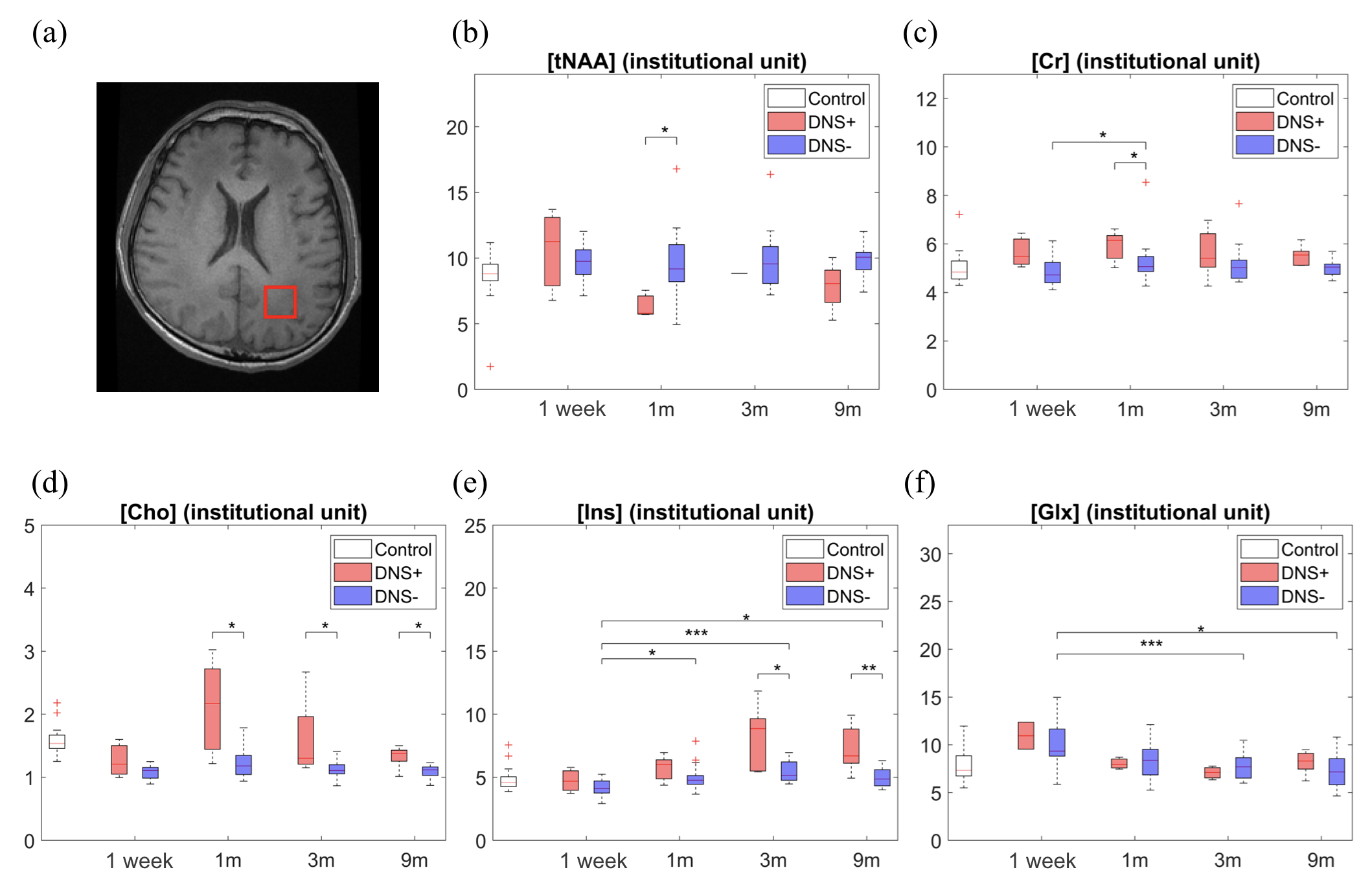
Figure 1.MRS voxel was placed at WM (red square) where WM abnormalities of DNS
frequently occurred in (a). (b)~(f): Box-and-Whisker plots of
absolute concentrations in institutional unit for tNAA, Cr, Cho, Ins, and Glx
among groups of normal subjects (Control), patients with (DNS+, red)) and
without DNS (DNS-, purple). Statistical
analysis with significance is indicated by '*' for p<0.05, '**' for p<0.01, ‘***’ for p<0.001.

Figure 2.Voxel-based DKI parameters (FA, AK, RK and MK)
were analyzed for 3 groups of WML+DNS+, WML+DNS- and WML-DNS-. Remarkable increase of AK, RK and MK at 1
month in patients with DNS (blue line) implies themselves as potential
indicators to discriminate between patients with and without DNS at early stage
of CO intoxication.
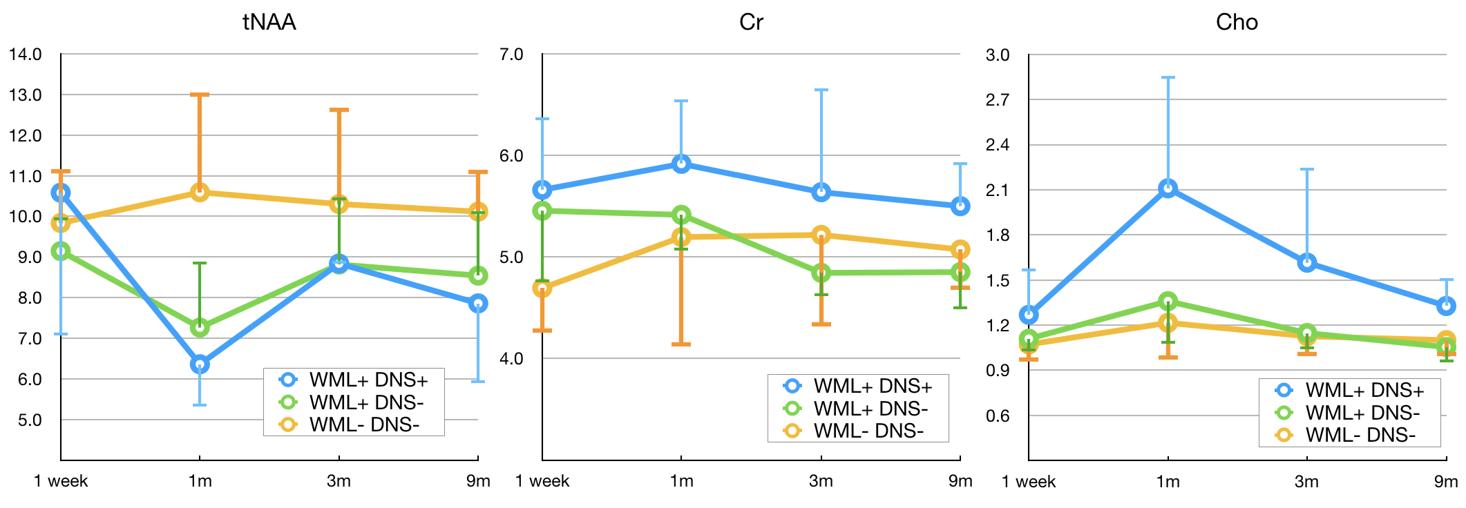
Figure 3.Absolute concentrations of tNAA, Cr and Cho in
mmol/mL (institutional unit) in patients of WML+DNS+ (blue), WML+DNS-(green)
and WML-DNS-(orange) at 1 week and 1, 3, and 9 months after CO intoxication. Cr
and Cho levels of DNS+ patients show significant increase especially at early
stage compared to DNS- patients either with or without WML. tNAA shows dramatical decrease for WML+
patients (blue and green) at 1 month and no significant difference between DNS+
and DNS- patients was found.
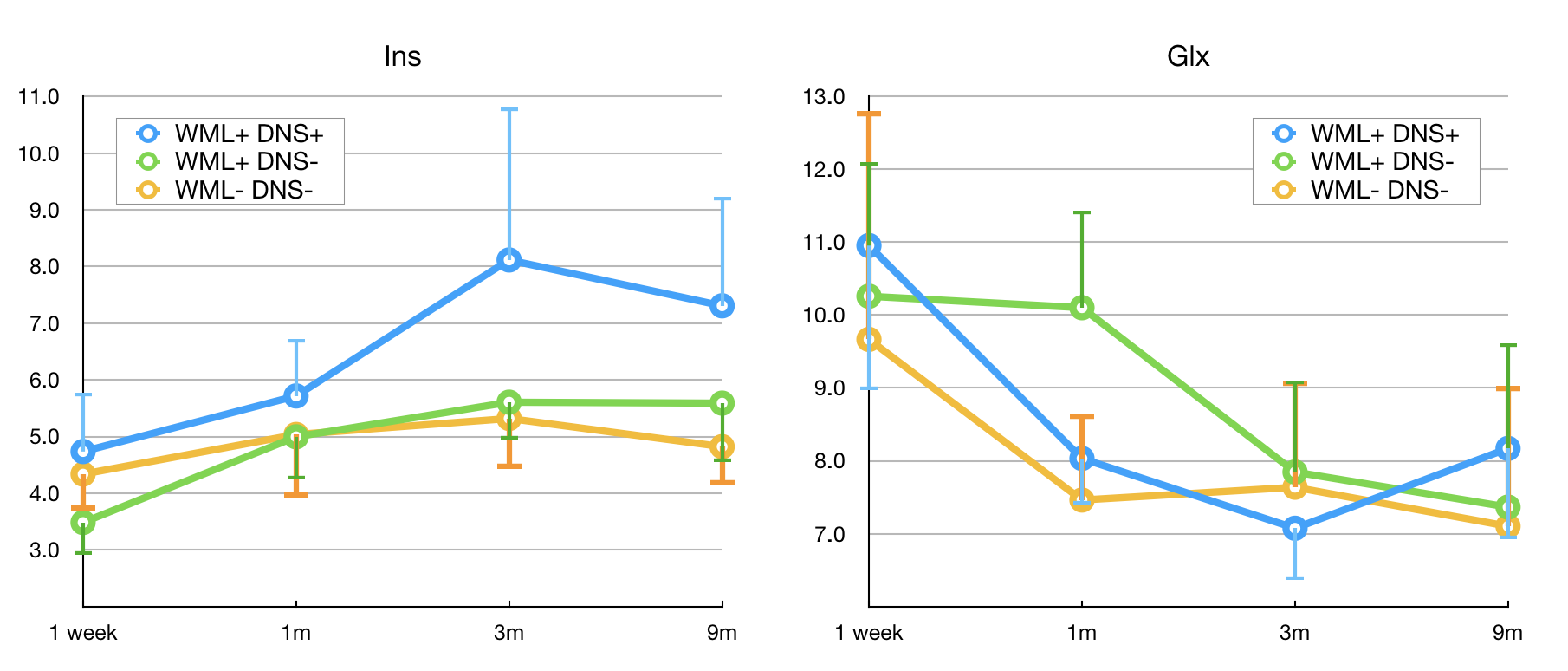
Figure 4.Absolute concentrations of Ins and Glx in
mmol/mL (institutional unit) in patients of WML+DNS+ (blue), WML+DNS-(green)
and WML-DNS-(orange) at 1 week and 1, 3, and 9 months after CO intoxication.
The longitudinal elevation of Ins level and descending of Glx in patients with
DNS suggest their association with neuropsychiatric activity and may help predicting prognosis of patients after CO intoxication.