2914
Over-discretized reconstruction to correct for B0 inhomogeneity and improve localization in 1H-MRSI of the prostate
Carlijn Tenbergen1, Loreen Ruhm2, Arend Heerschap1, Anke Henning2,3, and Tom Scheenen1
1Radiology and Nuclear Medicine, Radboud University Medical Center, Nijmegen, Netherlands, 2High-Field MR Center, Max Planck Institute for Biological Cybernetics, Tübingen, Germany, 3Advanced Imaging Research Center, University of Texas Southwestern Medical Center, Dallas, TX, United States
1Radiology and Nuclear Medicine, Radboud University Medical Center, Nijmegen, Netherlands, 2High-Field MR Center, Max Planck Institute for Biological Cybernetics, Tübingen, Germany, 3Advanced Imaging Research Center, University of Texas Southwestern Medical Center, Dallas, TX, United States
Synopsis
In in vivo MRSI spatial inhomogeneities of the main magnetic field cause spectral line broadening and location dependent frequency shifts in the spectra. An over-discretized reconstruction method is applied to existing prostate 1H-MRSI data, consisting of spatial over-discretization of the MRSI grid, frequency shift correction according to a high resolution B0map and weighted spatial averaging to the target resolution. Intravoxel B0 variations are corrected for and resampling to a new target resolution results in improved localization of signals. This could lead to detection of signals with lower spectral intensity and to improved visualization of smaller cancer lesions in the prostate.
Introduction
In in vivo magnetic resonance spectroscopic imaging (MRSI) different metabolic compounds of healthy or pathologic tissue, which are identified in a spectrum, can be spatially resolved in metabolite maps. With low spatial resolution and small matrix sizes, the spatial response function (SRF) of individual voxels extends all over the field of view, which can be reduced by hamming filtering of k-space at the cost of an even lower spatial resolution of the resulting metabolite maps. Another technical challenge in creating these maps is the spatial inhomogeneity of the main magnetic field (B0) across the organ of interest. Long-range B0 inhomogeneities introduced into voxel specific spectra by the SRF along with intra-voxel B0 inhomogeneity cause spectral line broadening and location dependent frequency shifts in the spectra. To reduce long range signal bleeding, to improve the effective versus the nominal voxel size and to correct for intra-voxel B0 inhomogeneity an over-discretized SRF target based reconstruction method1 is applied which uses high resolution B0 field maps2 to improve data quality of spectra and localization of the signals in metabolite maps derived from 1H-MRSI in the human prostate at 3T.Methods
The over-discretized reconstruction (ODR) method previously introduced for MRSI of the brain2 is applied to prostate 1H-MRSI data to assess feasibility. The method consists of two steps, the first of which generates a high resolution intermediate grid of subvoxels by over-discretizing the original MRSI dataset. Each subvoxel is then corrected with the frequency shift according to the local frequency offset from the center frequency of the main magnetic field at the specific location as indicated in a B0 map, acquired by a dual echo method3. As the spectral B0 correction is applied to the intermediate superresolution spectroscopic image, it allows to make use of the B0 map at high spatial resolution, correcting for (substantial) intravoxel variations. This causes metabolite peaks to align and removes noise correlations between neighbouring subvoxels. A weighted spatial average of subvoxels with now increased coherence, achieved by an SRF target that suppresses long range signal bleeds, leads to a final effective voxel size of the reconstructed MRSI data, very close to the original nominal voxel size.Prostate 1H-MRSI data, acquired with a semi-LASER 1H-MRSI sequence on a 3T MRI system (MAGNETOM Prisma-fit4, scan parameters: TR:750ms, TE:88ms, acquired voxelsize:7x7x7mm, varying matrix sizes:~11x11x11 voxels, 3 weighted averages and hamming filtering), is post-processed by the ODR. An over-discretization factor of 3 is chosen leading to an intermediate MRSI grid of 48x48 voxels. The B0map of the prostate is registered to and represented at the same resolution as the intermediate spectroscopic image. The corrected intermediate spectroscopic image is transformed to the target resolution of 16x16, by an SRF target with a characteristic width of 1.5 nominal voxels, consistent with the previous study.
The data processed with ODR and the originally acquired and postprocessed data (3D Hamming filter of zerofilled k-space) were imported in syngo.via4 and both fitted with the same prior knowledge. Metabolite maps of citrate and a combination of creatine, spermine and choline were created and signal to noise ratios (SNR) were extracted.
Results
Examples of spectra from voxels inside the peripheral zone of the prostate show similar or higher SNR for ODR data (Fig. 2). With the chosen parameters for over-discretization and subsequent averaging with the target function, the width of the targeted SRF at half height is 1.39 vs. 1.82 for the hamming-filtered data2. Therefore, effective voxels from ODR data are smaller, which is visible in citrate metabolite maps that adhere closer to the contours of the zone as depicted on the T2-weighted image (Fig. 3). High choline levels present in voxels matching the seminal vesicles is seen both in original and reconstructed data with less bleed of the signal further into the prostate in the latter dataset (Fig. 4).Discussion
The over-discretized reconstruction resulted in an improved localization of prostate metabolites in some subjects, showing a promising tool to increase spectral quality and spatial resolution of metabolite maps.Comparing SNR values between original and reconstructed data of the voxels within the VOI of the MRSI measurement is not straightforward. Voxels near the edges of the prostate, or within the transition zone, can contain less metabolite signal in the ODR data when compared to the original data with larger voxel size. Voxels completely within homogeneous peripheral zone tissue in both reconstructions do show an increase in SNR in the ODR case, even though the voxel size is smaller. A prerequisite for SNR gain for smaller voxels in ODR data seems to be good data quality. When the fitting protocol fails to correctly phase or identify peaks, or when steps in the B0 map of more than 2pi are present (and currently unaccounted for), the ODR algorithm is not able to increase spectral quality.
Conclusion
Over-discretized reconstruction of existing prostate 1H-MRSI data can lead to improved localization of metabolite signals. Currently there are some difficulties preventing the method to produce robust results for all spectra. When overcome, the method has potential to detect signals with lower spectral intensity and improve visualization of smaller cancer lesions in the prostate.Acknowledgements
No acknowledgement found.References
1. Kirchner, T. , Fillmer, A. , Tsao, J. , Pruessmann, K. P. and Henning, A. (2015), Reduction of voxel bleeding in highly accelerated parallel 1H MRSI by direct control of the spatial response function. Magn. Reson. Med., 73: 469-480.2. Kirchner, T. , Fillmer, A. and Henning, A. (2017), Mechanisms of SNR and line shape improvement by B0 correction in overdiscrete MRSI reconstruction. Magn. Reson. Med., 77: 44-56.
3. Schneider, E. and Glover, G. (1991), Rapid in vivo proton shimming. Magn. Reson. Med., 18: 335-347.
4. Siemens Healthcare GmbH, Erlangen, Germany
Figures
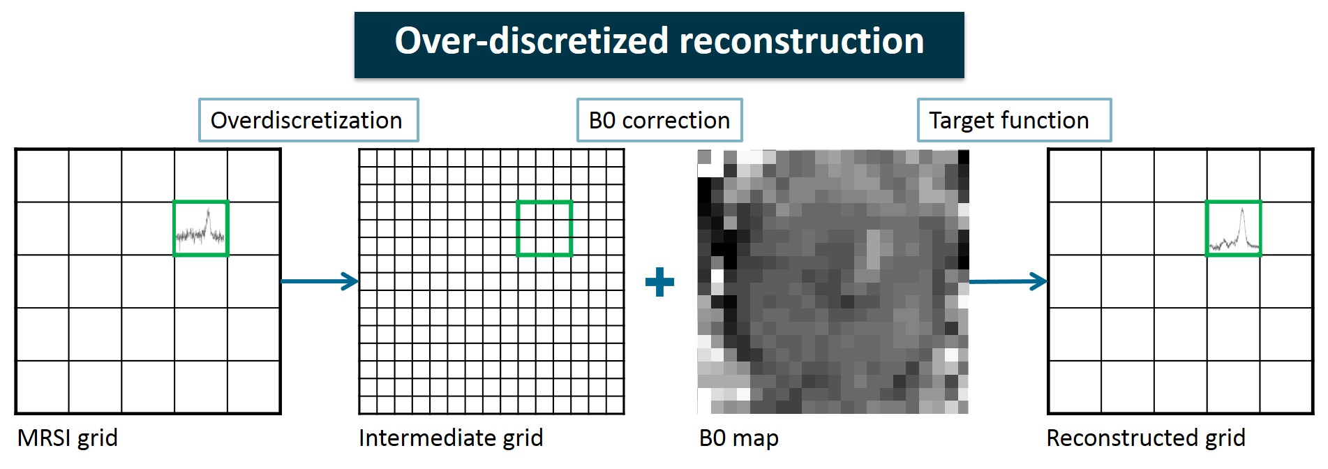
Figure 1: Schematic overview of the over-discretized reconstruction method. The original MRSI grid is first transformed into a higher-resolution intermediate grid by a factor 3. Each subvoxel spectrum is then frequency-shifted according to the corresponding B0map. By a weighted average of the subvoxels, the target resolution is achieved.
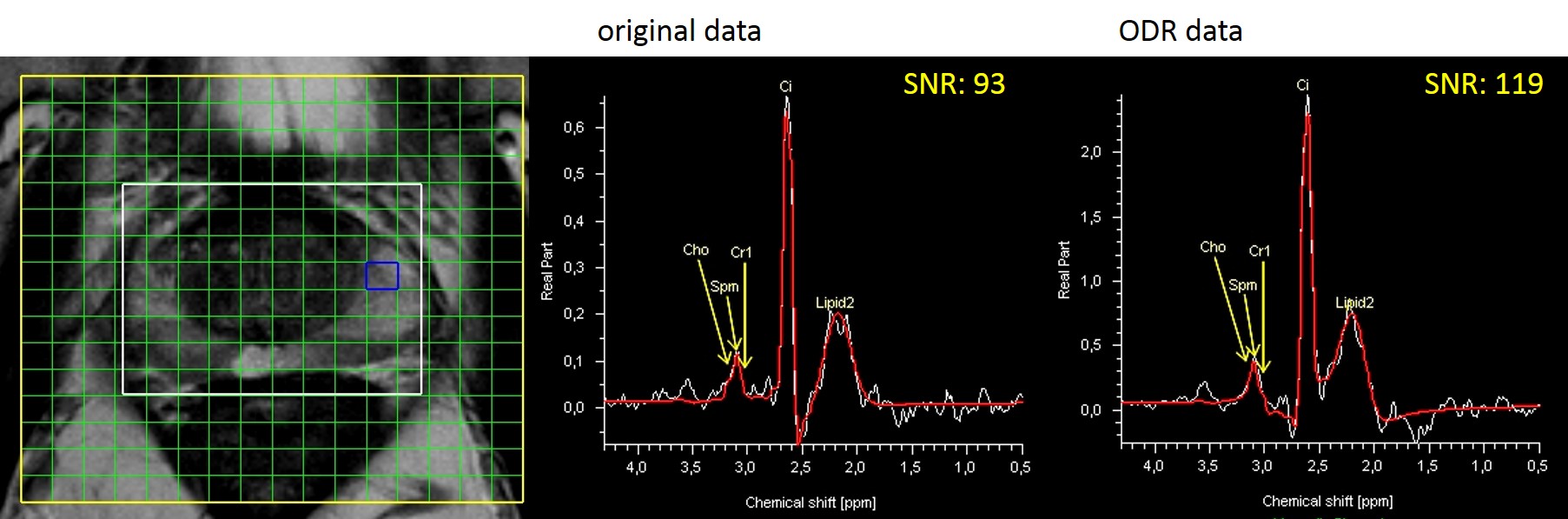
Figure 2: Two spectra (original hamming-filtered and ODR) fitted in syngo.via of the same location (centered around blue voxel) in the prostate of a patient. SNR values exported from syngo.via, with the citrate peak height as signal, show an increase by a factor of 1.3 for the ODR data (right panel) compared to the original data (middle panel).
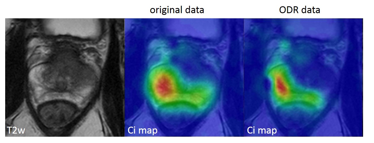
Figure 3: Improved localization in MRSI metabolite maps. In this subject the citrate signal is mostly found in the peripheral zone of the prostate. In the original dataset (middle panel) the Hamming filter induces bleed of the signal over the prostate. The dataset after ODR (right panel) shows the citrate signal with an improved localization to a specific spot in the peripheral zone with the signal adhering closer to the contours of the zone.
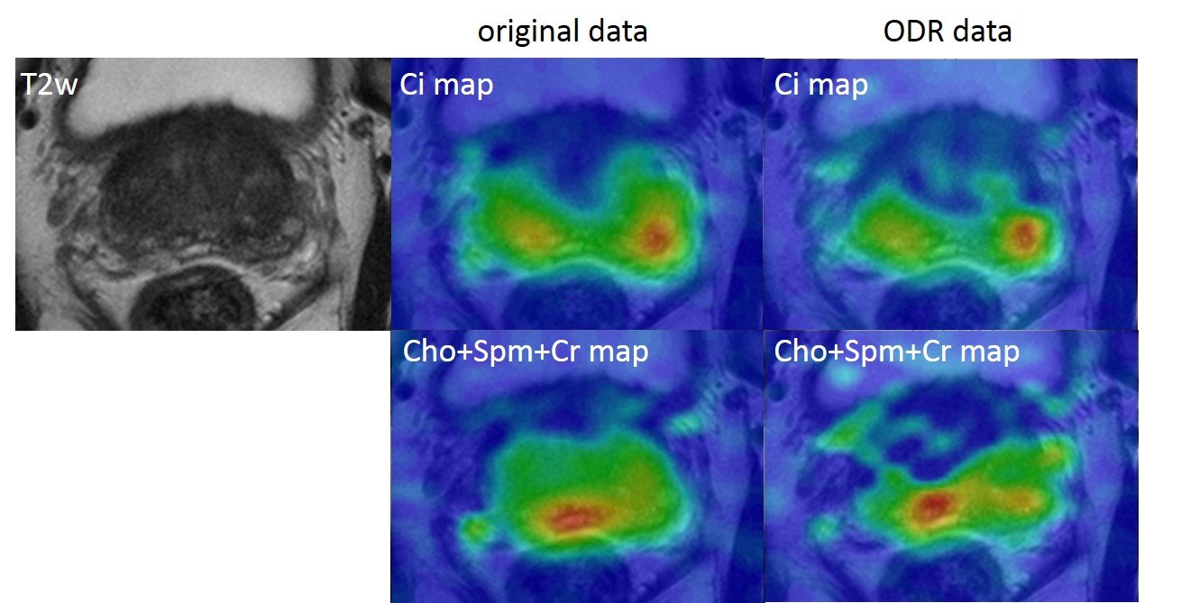
Figure 4: This
subject shows an improved localization of the citrate level to the
peripheral zone between original data (middle) and ODR data (right). The bottom
row shows high signal intensities arising from choline levels in the seminal vesicles, with improved localization for the ODR data in the right panel.