2878
Elliptical Localization with Pulsed Second-Order fields (ECLIPSE) for Lactate and BHB Edited Human Brain Proton MRSI1Radiology and Biomedical Imaging, Yale University, New Haven, CT, United States
Synopsis
Βeta-hydroxybutyrate (BHB) and lactate (Lac) are MRS visible metabolites important for brain energy metabolism. In this work we investigate the feasibility to detect elevated levels of BHB with proton MRSI following oral consumption of an energy drink containing 25g of a keto-ester. A gradient-modulated ECLIPSE-OVS method with minimal chemical shift displacement was implemented for lactate and BHB edited MRSI of the human brain. Elevated levels of BHB following oral intake of a keto-ester energy drink were observed in an 8 mL nominal volume MRSI acquired within 9-min at 4 T.
Introduction
Proton Magnetic Resonance Spectroscopic Imaging (MRSI) is a powerful technique that can spatially map the metabolic profile in the human brain, non-invasively1. The ability of MRSI to detect alterations in the neurochemical profile has seen applications in numerous brain pathologies.Βeta-hydroxybutyrate (BHB) and lactate (Lac) are MRS visible metabolites important for brain energy metabolism. Elevated levels of Lac have been reported in tumors, stroke, and hypoxia. BHB is one of three ketone bodies produced in the liver and serve as an alternate energy substrate during fasting or when on low carbohydrate diets (ketosis). Diet-induced ketosis has gained interest recently2,3 as a treatment option for brain pathologies, such as tumors, epilepsy, and neurodegenerative diseases.
While MRSI is a potentially powerful technique, it demands high magnetic field homogeneity, water and especially lipid suppression. Elliptical localization with pulsed second order fields (ECLIPSE4) has recently been proposed as a novel method for robust (> 100-fold) lipid suppression with high brain coverage and low RF power requirements.
In this work we investigate the performance of a gradient-modulated ECLIPSE-OVS method for Lac and BHB edited MRSI, and investigate the feasibility to detect elevated levels of BHB following oral consumption of a keto-ester energy drink.
Methods
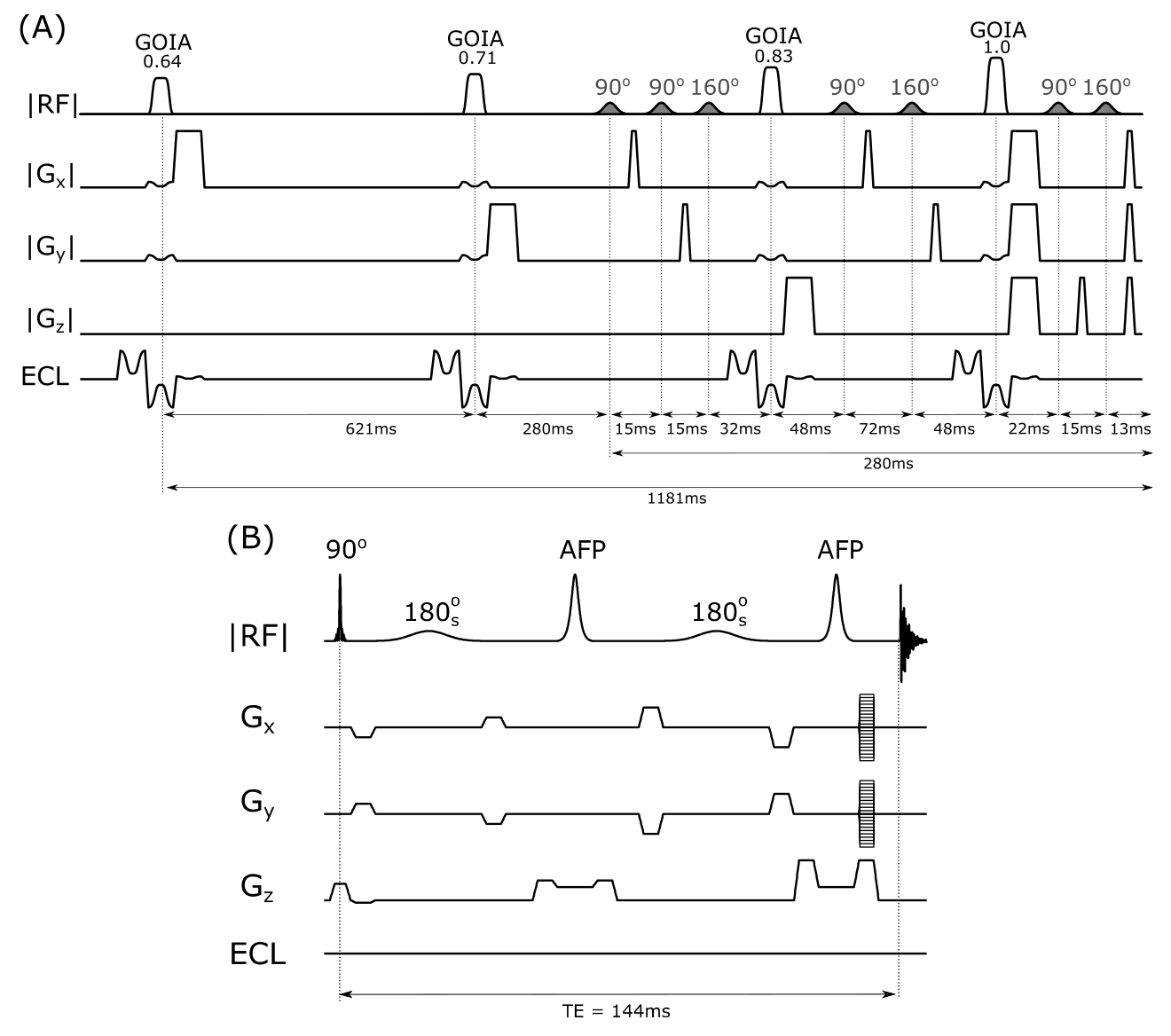
All MR experiments were performed on a 4T magnet (Magnex Scientific Ltd.) interfaced to a Bruker Avance III HD spectrometer running ParaVision 6 (Bruker, Billerica, MA, USA). A within-brain B1+ optimized, 8-element Tx/Rx volume coil embedded in the ECLIPSE gradient coil was used for the study in a fixed phase configuration. The ECLIPSE system4 is a home-built, unshielded gradient insert consisting of Z2, X2Y2, and XY second order spherical harmonic magnetic fields with efficiencies of 5.48, 2.58 and 2.76 Hz/cm2/A, respectively, interfaced to a home-built multi-channel gradient controller5. ECLIPSE-OVS is achieved by repeatedly exciting and dephasing an elliptical ROI. Due to the absence of overlapping slices, the optimization of nutation angles and delays with ECLIPSE is straightforward. In order to minimize chemical shift displacement, each ECLIPSE-OVS pulse was executed with gradient-modulation based on the GOIA algorithm6 (6.66ms, 15 kHz BW), derived from an AFP-HS4 pulse (6.66ms, 3 kHz BW). A four-RF pulse-based ECLIPSE-OVS module was developed (Figure 1A). A seven-pulse VAPOR-style water suppression sequence was optimized using Gaussian pulses (10ms, 200Hz BW) and interleaved within the ECLIPSE-OVS module.The MRSI component was achieved with an AFP double spin-echo with phase encoding gradients following the final AFP pulse (Figure 1B). The editing pulses were set at 4.1 ppm, and +1000Hz for edit ON and edit OFF scans, respectively. Constant-field B0 eddy currents associated with unshielded ECLIPSE Z2 pulses were minimized by having -109% and -9% pre and post-pulses. No further eddy current compensations were performed. The overall ECLIPSE-OVS based MRSI sequence TE/TR was 144ms/2000ms.
BHB and Lac-edited MRSI scans were acquired before and circa 85-min following the oral administration of 25 g of a keto-ester energy drink (HVMN, San Francisco, CA, USA). The blood ketone levels increased from ~0.1 mM to ~3.6 mM circa 45-min after keto-ester consumption.
Results
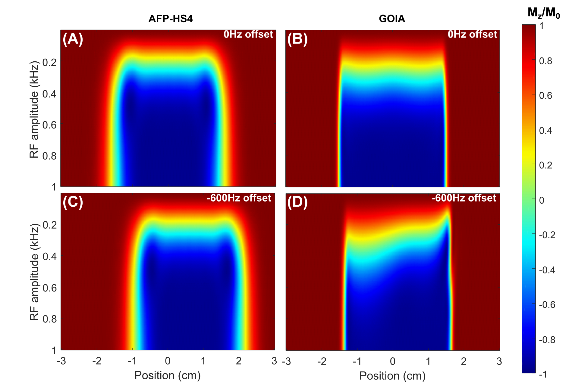
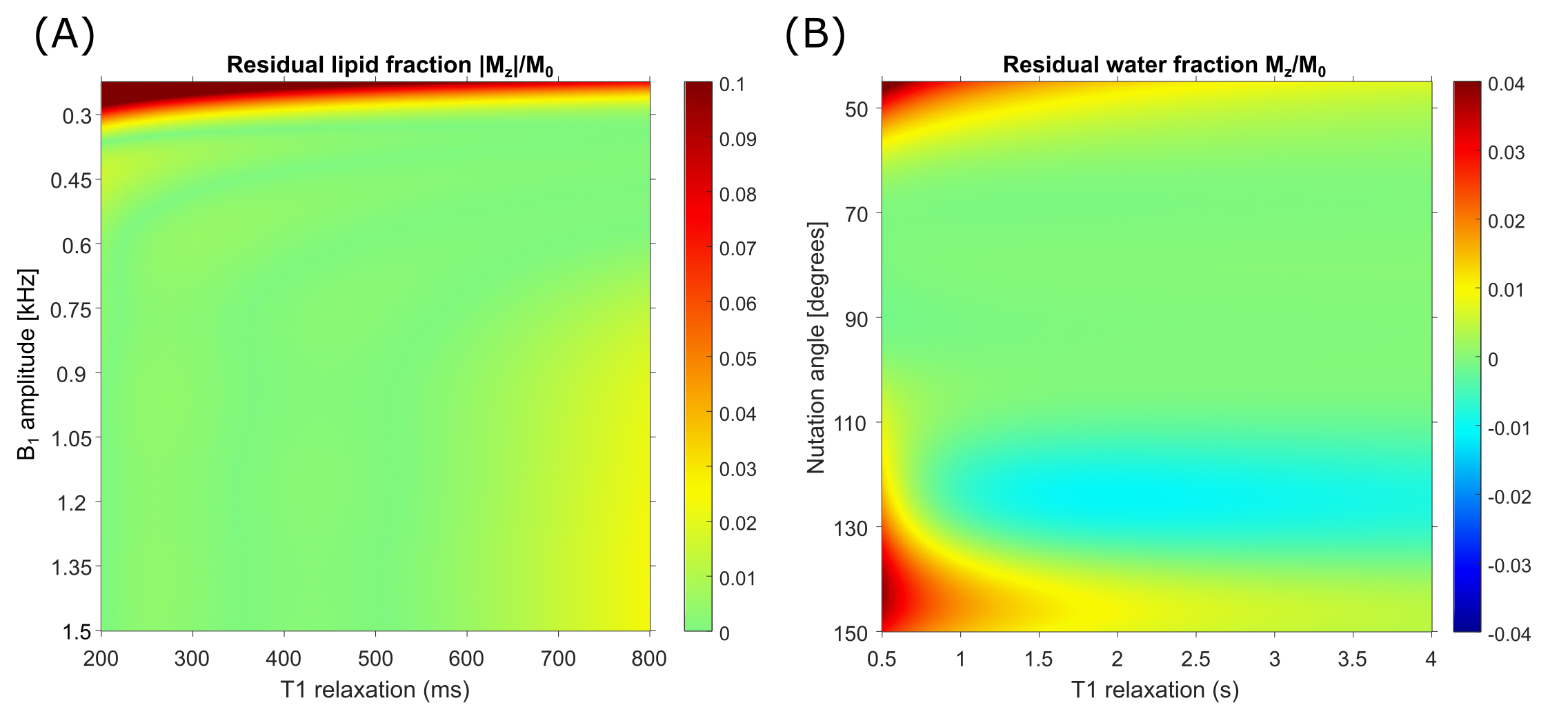
Bloch simulations for the AFP-HS4 pulse and GOIA pulse, vs B1 amplitude are illustrated in Figure 2A-B. The simulation is repeated for a -600Hz frequency offset (fat-water chemical shift at 4T), as illustrated in Figure 2C-D. GOIA-based OVS provides a smaller chemical shift displacement (1.2 mm) than AFP-HS4 pulse based OVS (6 mm). However, at low RF amplitude the GOIA profile gives an asymmetric frequency profile. While this could be compensated for by ~26% higher B1 amplitude, a more power-efficient solution was to execute GOIA-OVS on-resonance for lipid resonances at ~1.2 ppm.Lipid suppression of the optimized GOIA-ECLIPSE-OVS module as a function of T1 and B1 is illustrated in Figure 3A. Water suppression performance in simulation, as a function of T1 and B1 is shown in Figure 3B.
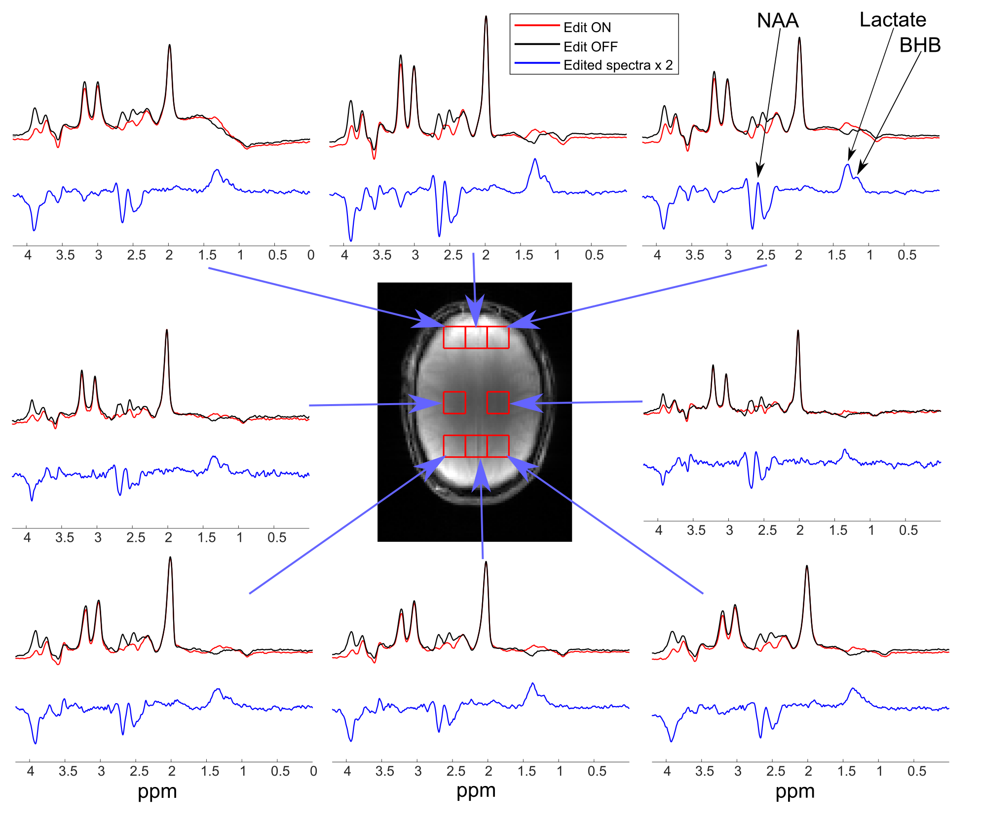
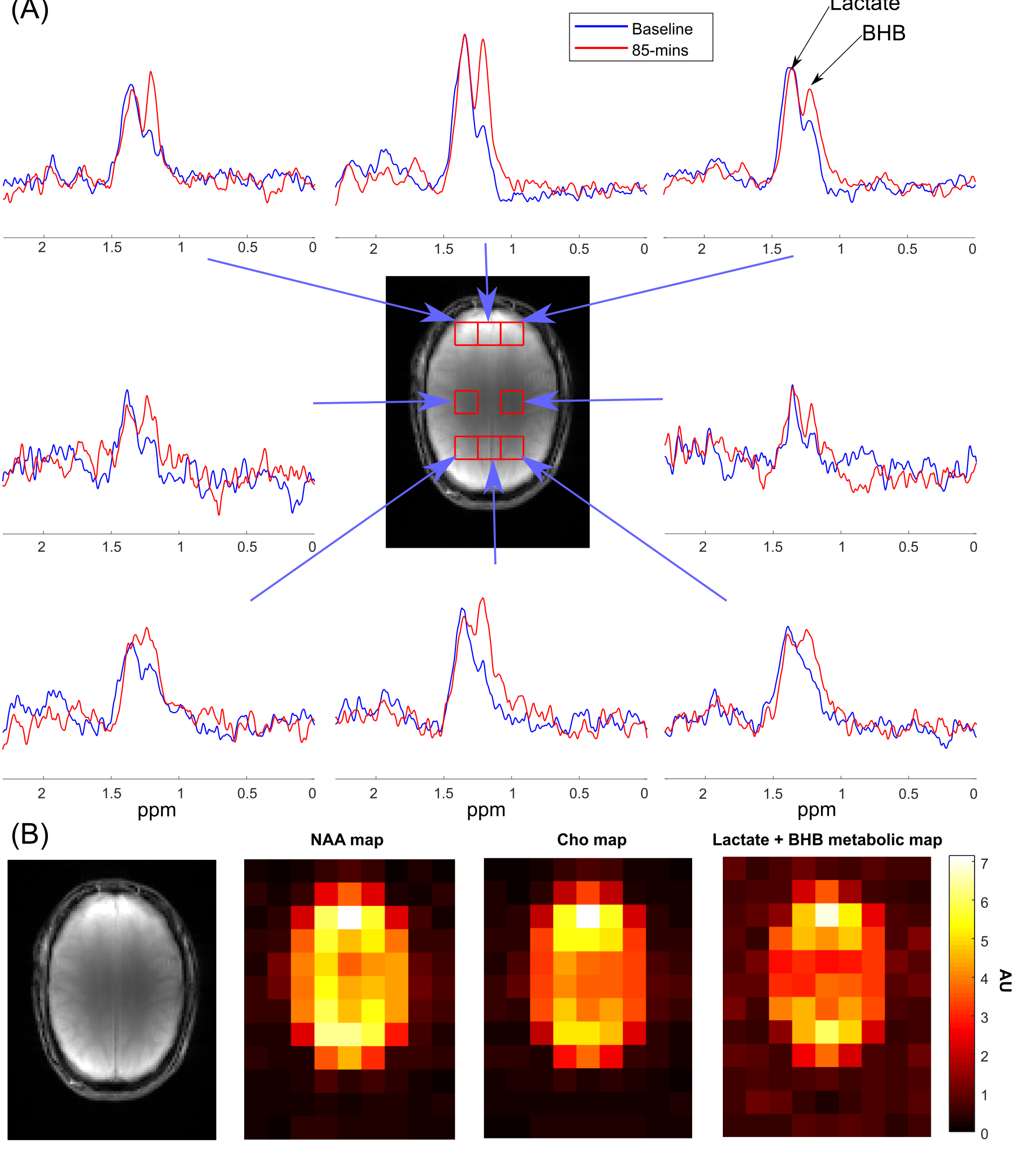
Figure 4 illustrates edit-ON (red), edit-OFF (black), and edited (blue) spectra for eight voxel locations, acquired from a healthy volunteer. In all edited spectra lactate at 1.31 ppm is present, with a lower amplitude BHB peak at 1.19ppm. Figure 5A illustrates a comparison of edited spectra from the baseline scan and 85-mins post keto-ester consumption, where the BHB signal is noticeably elevated.
Effective extracranial lipid suppression was present in both scan sessions, as is evident from NAA, and creatine metabolic maps. Lactate + BHB metabolic maps were generated from the edited MRSI data (Fig. 5B), and were also free from extracranial lipid signals.
Conclusions
An ECLIPSE-OVS method was developed with gradient-modulated GOIA RF pulses for lactate and BHB edited MRSI of the human brain. The gradient-modulation provides a five-fold improvement in chemical shift displacement with an RF peak power increase of ~21%, and an average RF power increase of 36% relative to the AFP HS4 pulse. Elevated levels of BHB following oral intake of a keto-ester were seen across the brain in 9-min of MRSI acquisition. GIOA-based ECLIPSE-OVS provides high-quality lipid suppression with excellent elliptical brain coverage that can benefit a wide range of MRSI applications. The low RF power requirements in combination with the high bandwidth will be beneficial for applications at 7 T.Acknowledgements
This research was supported by NIH grant R01- EB014861, and a James S. McDonald Foundation planning grant.References
[1] Maudsley AA, Domenig C, Govind V, Darkazanli A, Studholme C, Arheart K, Bloomer C. Mapping of brain metabolite distributions by volumetric proton MR spectroscopic imaging (MRSI). Magn Reson Med. 2009; 61:548-559.
[2] Weber D, Aminazdeh-Gohari S, Kofler B. Ketogenic diet in cancer therapy. Aging. 2018; 10(2):164-165.
[3] Branco A, Ferreira A, Simoes R, et al., Ketogenic diets: from cancer to mitochondrial diseases and beyond. Eur J Clin Invest. 2016; 46(3):285-298.
[4] de Graaf RA, Brown PB, De Feyter HM, McIntyre S, Nixon TW. Elliptical localization with pulsed second-order fields (ECLIPSE) for robust lipid suppression in proton MRSI. NMR in biomedicine 2018;31(9):e3949.
[5] Nixon TW, McIntyre S, de Graaf RA. The design and implementation of a 64 channel arbitrary gradient waveform controller. Proc Int Soc Magn Reson Med. 2017;25:969.
[6] Tannus A, Garwood M. Adiabatic pulses. NMR in biomedicine 1997;10(8):423–434.
Figures