2875
Development of hydroxyl radical imaging using dynamic nuclear polarization MRI1Gifu University, Gifu, Japan, 2Kyushu University, Fukuoka, Japan
Synopsis
Oxidative stress is implicated in various diseases such as inflammation, neurodegenerative disorders (Alzheimer’s disease, Parkinson’s disease), atherosclerosis, diabetic and cancer. Excess reactive oxygen species (ROS) are produced during altered cellular metabolism in various diseases. Among ROSs, hydroxyl radicals (•OH) is one of the most reactive molecules in biological systems. Therefore, it is considered that monitoring of •OH is could be useful technologies for evaluation of redox mechanism and oxidative diseases. In this study, we developed the hydroxyl radical imaging technique using combining DNP-MRI and DMPO.
INTRODUCTION
Oxidative stress is implicated in various diseases such as inflammation, neurodegenerative disorders (Alzheimer’s disease, Parkinson’s disease), atherosclerosis, diabetic and cancer1. Excess reactive oxygen species (ROS) are produced during altered cellular metabolism in various diseases2. It is known that ROS can play both beneficial and harmful roles in the physiology of cells. For example, ROSs are acting as messengers in cell-signaling pathways, on the other hand, the harmful effects of ROS include oxidative damage to biomolecules, such as lipids, proteins, nucleic acids and sugars. Among ROSs, hydroxyl radicals (•OH) is one of the most reactive molecules in biological systems3. Therefore, it is considered that monitoring of •OH is could be useful technologies for evaluation of redox mechanism and oxidative diseases.The electron paramagnetic resonance (EPR) spectroscopy with spin trap agent is a convenient and popular technique to identify the ROS. With this technique, short-lived oxygen derived radicals are trapped by a spin trap agent to form long-lived radicals4. The most popular spin trap agent is 5,5-dimethyl-1-pyrroline N-oxide (DMPO), and spin adduct of DMPO with hydroxyl radical gives hydroxyl radical-specific EPR spectrum as DMPO-OH5. Therefore, imaging of DMPO-OH free radical might be useful methods for clarify the hydroxyl radical generation and examine the effect of antioxidant capability.
In vivo Dynamic nuclear polarization (DNP)-MRI is a noninvasive imaging method to obtain the spatio-temporal information of free radicals with MRI anatomical resolution6. The proton signal in tissues including free radicals as DNP effect can be dramatically enhanced by EPR irradiation at the resonance frequency of the free radical prior to applying the MRI pulse sequence. In this study, we developed the hydroxyl radical imaging technique using combining DNP-MRI and DMPO.
METHODS
The Fenton reaction (H2O2 and FeSO4.) was utilized for hydroxyl radicals and DMPO was selected as the spin trap agent for EPR spectroscopy. Hydroxyl radical generation as DMPO-OH signal was confirmed by EPR spectroscopy. For DNP-MRI measurement, a two-tube phantoms (200 μL, 5.4 mm deep, 9 mm long) were prepared for comparison of the DNP phenomenon. In one tube, H2O2 and FeSO4 were mixed and DMPO was added just before the DNP measurement. Another one was filled with PBS. A surface coil for EPR irradiation was utilized for DNP-MRI measurements. In order to determine the optimal EPR excitation frequency of DMPO-OH in DNP-MRI, DNP-MRI measurements at various EPR frequency from 460 to 483MHz were performed. In addition, to determine the suitable concentration of hydrogen peroxide, DNP-MRI with DMPO measurement were performed using various concentration of hydroxyl radical. The scanning conditions for the DNP-MRI experiment was follows: power of EPR irradiation = 7 W, flip angle = 90°, repetition time (TR) × echo time (TE) × TESR = 500 × 25 × 250 ms, number of acquisitions = 10 and number of phase-encoding steps = 32. Field of view (FOV: 40 × 40 mm) was represented by a 64 × 64 matrix after image reconstruction.RESULTS and DISCUSSION
At first, we confirmed the EPR spectrum of DMPO-OH by Fenton reaction between H2O2 and FeSO4. The EPR spectrum was stably observed until 10 min after generation of hydroxyl radical.In DNP-MRI measurement, it is important to determine the suitable EPR irradiation frequency to obtain the maximum effect of DNP. We confirmed the concentration of FeSO4 (4mM) used in this study is not affected on DNP-MRI signal. We measured phantoms on various EPR irradiation frequencies (1 MHz interval from 460 to 483MHz) using DNP-MRI. DNP enhancement was observed from 471 MHz and maximum enhancement was observed at 474.5MHz. The image intensity of DMPO-OH on EPR ON was 3 times higher than that of EPR OFF image. On the other hand, there was no enhancement in the PBS tube. Next, we changed the H2O2 concentration to observe the hydroxyl radical dependent DNP enhancement. DNP enhancement was clearly increased depending on H2O2 concentration. From these experiments, dose dependency of hydroxyl radical generation as a DMPO-OH could be detected by DNP-MRI system.
H2O2 has been utilized as a radio-sensitizer on Kochi Oxydol Radiation Therapy for Unresectable(KORTUC) treatment for cancer patients7. Because the distribution of H2O2 is important for treatment efficacy, visualization of H2O2 derived hydroxyl radical might be useful for determination of suitable administration method on KORTUC treatment. In addition, this technology could be utilized in vivo redox monitoring on various oxidative disease although the higher sensitivity and stability for spin trapping monitoring by DNP-MRI is required.
CONCLUSION
In this study, we observed the DNP enhancement derived from DMPO-OH by hydroxyl radical generation system and succeeded in visualization of DMPO-OH by DNP-MRI. In addition, we determined the optimal EPR irradiation frequency for DMPO-OH in DNP-MRI system.Acknowledgements
No acknowledgement found.References
1. Termini J. Hydroperoxide-induced DNA damage and mutations. Mutat Res 2000;450:107–24.
2. Aleksandra K, Luminescence. Studies on the antioxidant activities of some new chromone compounds. Luminescence. 2014 Nov;29(7):846-53.
3. Toyokuni S. Role of iron in carcinogenesis: cancer as a ferrotoxic disease. Cancer Sci 100:9–16.
4. Nakagawa S. Estimation of Relative Reaction Rate of Hydroxy Radical with Poly-hydroxy Benzenes: ESR Spin Trapping Combined with UV-A Photolysis. Anal Sci. 2013;29(3):377-80.
5. Ranguelova K, Mason RP. The Fidelity of Spin Trapping with DMPO in Biological Systems. Magn Reson Chem. 2011 Apr;49(4):152-8.
6. Hyodo F, Naganuma T, Eto H, et al. In vivo melanoma imaging based on dynamic nuclear polarization enhancement in melanin pigment of living mice using in vivo dynamic nuclear polarization magnetic resonance imaging. Free Radic Biol Med. 2019 Apr;134:99-105.
7. Ogawa Y, Ue H, Tsuzuki K, et al. New radiosensitization treatment (KORTUC I) using hydrogen peroxide solution-soaked gauze bolus for unresectable and superficially exposed neoplasms. Oncol Rep. 2008 Jun;19(6):1389-94.
Figures
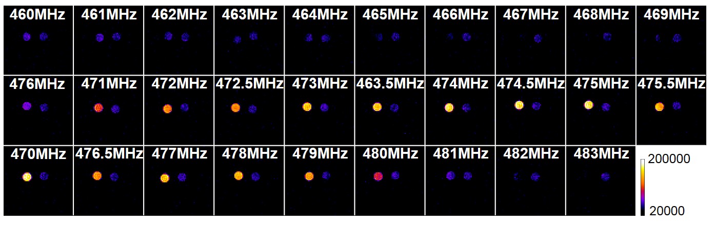
Fig1. Determination of the suitable EPR irradiation frequency for DMPO-OH using DNP-MRI. DNP-MRI measurements were performed using various EPR frequency from 460MHz to 483MHz. The DNP enhancement by DMPO-OH was observed and the highest enhancement was obtained at 474.5 MHz.