2842
Initial Assessment of a novel hybrid ultrashort echo time (UTE) sequence
Lumeng Cui1, Emily J McWalter1,2, Gerald Moran3, and Niranjan Venugopal4
1Division of Biomedical Engineering, University of Saskatchewan, Saskatoon, SK, Canada, 2Department of Mechanical Engineering, University of Saskatchewan, Saskatoon, SK, Canada, 3Siemens Healthcare Limited, Oakville, ON, Canada, 4Department of Radiotherapy Physics, CancerCare Manitoba, Winnipeg, MB, Canada
1Division of Biomedical Engineering, University of Saskatchewan, Saskatoon, SK, Canada, 2Department of Mechanical Engineering, University of Saskatchewan, Saskatoon, SK, Canada, 3Siemens Healthcare Limited, Oakville, ON, Canada, 4Department of Radiotherapy Physics, CancerCare Manitoba, Winnipeg, MB, Canada
Synopsis
Ultrashort echo time (UTE) pulse sequences have the unique ability to visualize short T2 tissues. While several UTE techniques have been implemented and demonstrated good clinical results, there currently does not exist a flexible and robust UTE pulse sequence which can easily manipulate multiple parameters “on-the-fly“ (i.e. selection of trajectories, excitation pulses, acquisition parameters, etc.), and obtain several complimentary scans in a single scan session. In this work we present a newly developed hybrid UTE pulse sequence that includes multiple excitation pulses, and varying trajectories, allowing for novel investigations in studying short T2 tissues.
Introduction
Short T2 tissues appear as a signal void on conventional MRI images due to their short relaxation times1; however, there are several applications in which signal is required from these tissues. Signal can be obtained from short T2 tissues using Ultrashort Time-of-Echo (UTE) sequences2 that employ approaches such as non-Cartesian trajectories and a half-pulse or hard pulse to reduce TE to the system limits2-5. Tissues such as bone, tendons, ligaments and deep layers of cartilage have been successfully visualized using UTE approaches however it is not clear what particular trajectories, pulses, and associated parameters are optimal for this visualization. This is because a single sequence for direct comparisons is lacking. Thus, presented in this study, is a flexible robust UTE sequence that incorporates varying combinations of trajectories, pulses, and voxel sizes which enable multiple UTE data acquisitions in a single scanning session.Methods
A flexible UTE sequence was developed (and tested on a 3T MRI (MAGNETOM Skyra, VE11C, Siemens Healthcare, Erlangen, Germany) using an in-house phantom that contained both short and long T2 properties. The current sequence can generate on-the-fly, radial and spiral trajectories using the inline GUI. 2D acquisitions were developed using radial and spiral trajectories and employed a half-Sinc excitation pulses. Similarly, 3D acquisitions were developed using radial and spiral trajectories, but have the option of using a half-Sinc or hard excitation pulses. The images were reconstructed using standard methods. To measure the limits of the sequence, and its execution on the 3T MRI, data was collected by 1) Varying the numbers of spokes/interleaves and cones that comprised the radial and spiral trajectories, 2) Performing an ultrashort and a long TE acquisitions, and 3) Switching between the use of the half-Sinc versus hard pulses (See details in Figure 1). To evaluate the acquired data, we observed the variation of the image artifacts for 1), conducted a dual-echo subtraction (ultrashort and long) for 2), and reported Signal-to-Noise-Ratio (SNR) in Figure 5.b using the two-region method6 for 3).Results
When the number of shots was increased, the artifacts observed in fewer shots start disappearing for radial and spiral trajectories in the 2D acquisition (Figure 2.a-n); and their results at a high shot number (96/1024 shots for spiral/radial) are comparable to the GRE image. In the 3D spiral-cone trajectory, when the cone number is increased, the image quality improves with less artifacts (Figure 3.a.1-6). In terms of spoke-cone combination, 512*256 or 362*362 suggests a better image quality than other combinations (Figure 3.b.1-8 and c.1-8). With both the radial and spiral trajectories it is feasible to visualize short T2 components (Figure 4.a-f). When the half-Sinc and the Hard pulses were compared the spiral trajectories generally had higher SNR (Figure 5.a.1-8 and b).Discussion
We showed that it was possible to develop and use a flexible UTE sequence to image short T2 substances. When comparing the radial and spiral trajectory (Figure 2.a-n), the undersampled shots result in the undersampling artifact for radial trajectory; while for spiral trajectory, long spiral gradient readout due to fewer shots cause the off-resonance artifact as observed. However, increasing the number of shots can effectively suppress the artifacts, and the images acquired with a high shot number are comparable to the GRE image while achieving an ultrashort TE (0.1 – 1 ms1). In a 3D trajectory, the artifacts are also from fewer cones in addition to fewer spokes/interleaves. For example, with an insignificant off-resonance artifact (Figure 3.a.1-6), the artifacts are successfully reduced from 32 cones to 512 cones. Even though a larger number of spokes and cones are more desirable for the 3D radial to obtain a high-quality image, there is a limit for the number of gradients that the MR system can execute. Therefore, we need to determine an optimal combination between spokes and cones (Figure 3.b.1-8 and c.1-8); the results suggest that spokes have more impact on the image quality than the cone number. 3D radial and spiral can both capture more short-T2 signals with an ultrashort TE (0.06 ms) than a longer TE (3.09 ms) (Figure 4); the short T2 details (arrows) that are observed as voids in a long TE image can be further enhanced by taking the subtraction between two TEs. For the radial trajectory, the half-Sinc pulse’s SNRs on short T2, long T2, and the whole region are higher than the SNRs of the hard pulses (with different flip angles), but the total acquisition time of half-Sinc excitation is twice that of the hard pulse excitation (Figure 5). For the spiral trajectory, there is an evident difference of SNR on the short T2 and whole region between the half-Sinc pulse and hard pulses, and the hard pulses with high flip angles demonstrate a higher SNR on the long T2 region.Conclusion
The newly developed hybrid UTE sequence demonstrated efficacy in preliminary phantom tests. Additionally, the new sequence provided a flexible platform to adapt the pulse sequence for a variety of UTE applications by enabling the end-user to employ varying trajectories, dimensions, and excitation pulses.Acknowledgements
No acknowledgement found.References
- Bydder GM. The Agfa Mayneord lecture: MRI of short and ultrashort T 2 and T 2* components of tissues, fluids and materials using clinical systems. The British journal of radiology. 2011 Dec;84(1008):1067-82.
- Gold GE, Pauly JM, Macovski A, Herfkens RJ. MR spectroscopic imaging of collagen: tendons and knee menisci. Magnetic resonance in medicine. 1995 Nov;34(5):647-54.
- Bergin CJ, Pauly JM, Macovski A. Lung parenchyma: projection reconstruction MR imaging. Radiology. 1991 Jun;179(3):777-81.
- Glover GH. Simple analytic spiral k‐space algorithm. Magnetic Resonance in Medicine: An Official Journal of the International Society for Magnetic Resonance in Medicine. 1999 Aug;42(2):412-5.
- Cline HE, Zong X, Gai N. Design of a logarithmic k‐space spiral trajectory. Magnetic Resonance in Medicine: An Official Journal of the International Society for Magnetic Resonance in Medicine. 2001 Dec;46(6):1130-5.
- Dietrich O, Raya JG, Reeder SB, Reiser MF, Schoenberg SO. Measurement of signal‐to‐noise ratios in MR images: influence of multichannel coils, parallel imaging, and reconstruction filters. Journal of Magnetic Resonance Imaging: An Official Journal of the International Society for Magnetic Resonance in Medicine. 2007 Aug;26(2):375-85.
Figures
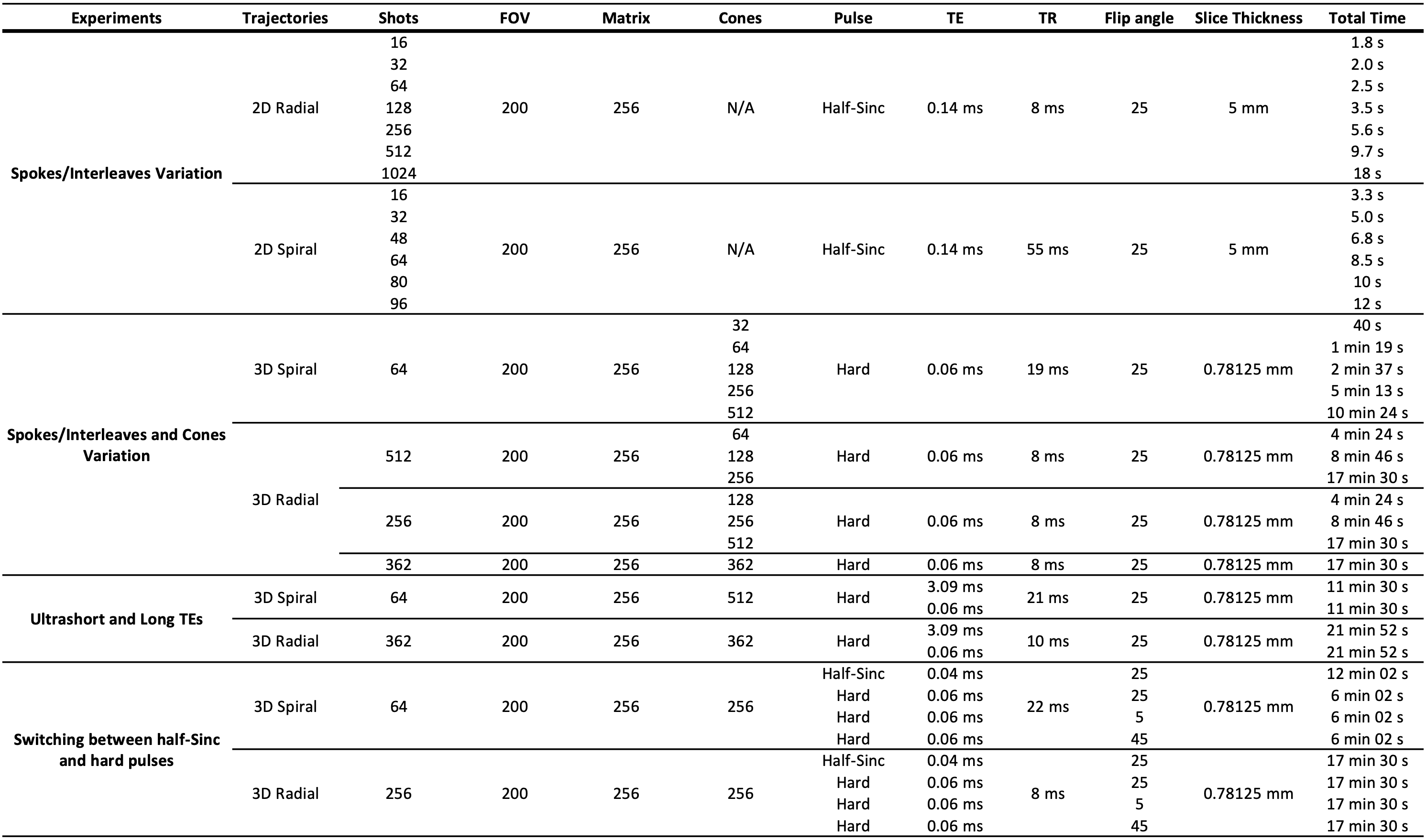
Figure 1. Experiments setups and protocols for 1) Varying the numbers of spokes/interleaves and cones that comprised
the radial and spiral trajectories, 2) Performing an ultrashort and a long TE
acquisitions and 3) Switching between the use of the half-Sinc versus hard
pulses.
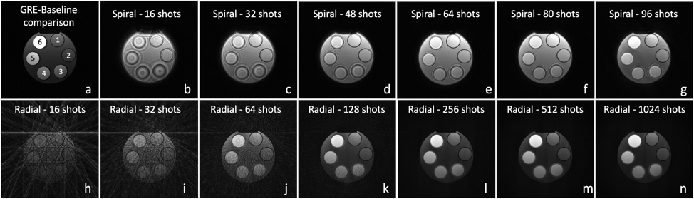
Figure 2. (a) GRE,
(b-g) 2D spiral (TE 0.14 ms and TR 55 ms), and (h-n) 2D Radial
(TE 0.14 and TR 8 ms) acquisitions. Label 1-6 in GRE indicate the property of the
phantom from a short-T2 to a long-T2.
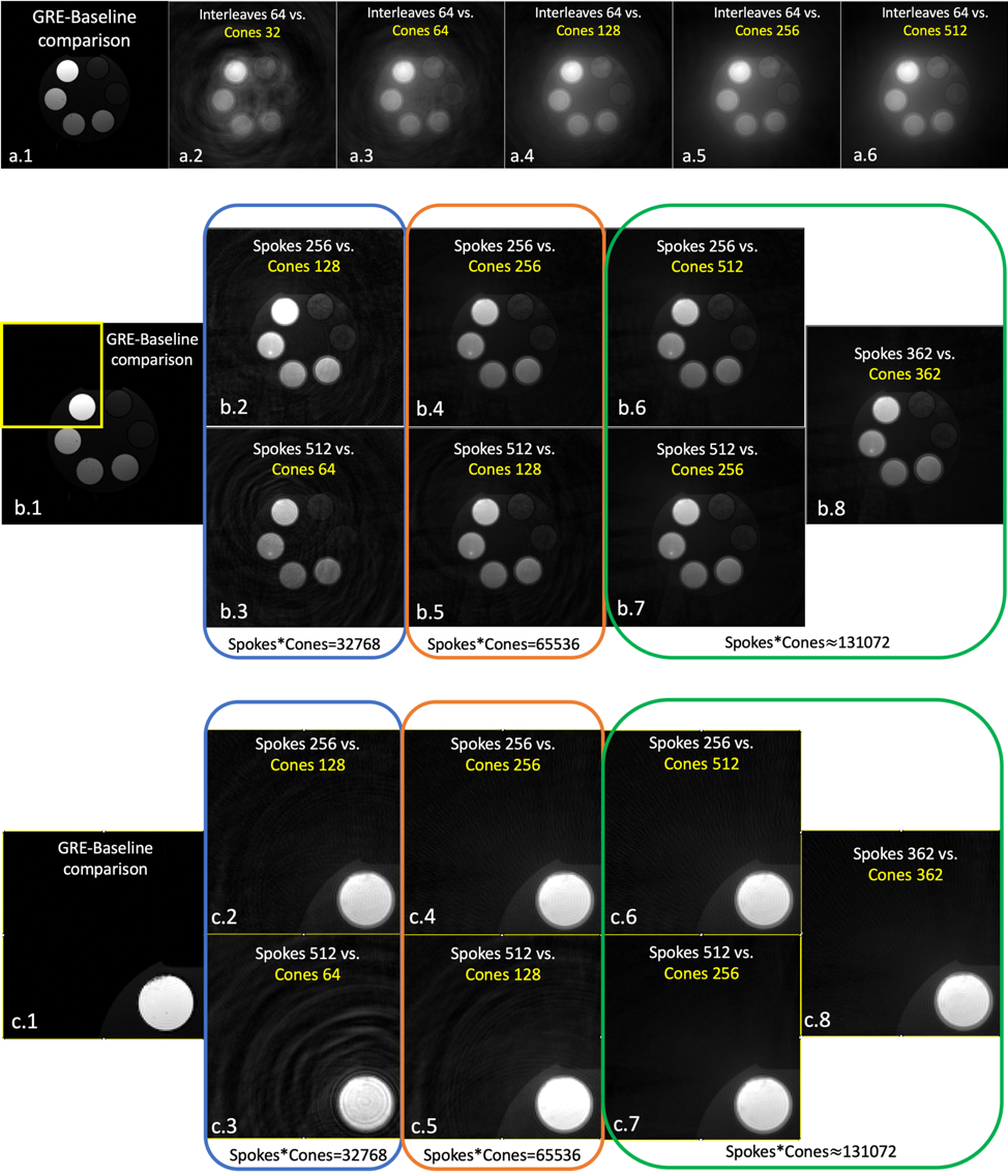
Figure 3. (a.1)
GRE-baseline comparison, (a.2-6) 3D Spiral acquisitions with a vary of
cone number; (b.1) GRE-baseline comparison with an indication
enlargement box, (b.2-8) 3D Radial acquisitions with different
combinations of cones and spokes comparing; (c.1) The enlargement of
GRE-baseline comparison, (c.2-8) Enlargement of (b.2-8).
The image quality can be improved with less artifacts by varying the numbers of spokes/interleaves and cones that
comprised the radial and spiral trajectories.
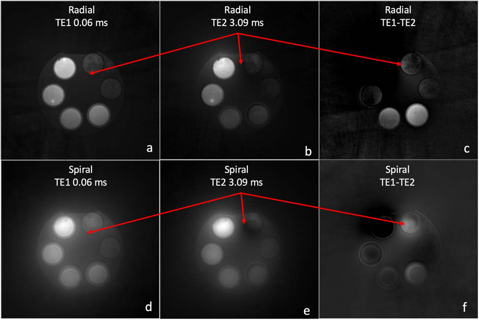
Figure 4. Short T2 substances imaging of
3D radial (a-c) and spiral (d-f)
trajectories. The arrows point at the short T2
contrast on a short TE, a long TE, and TE difference images.
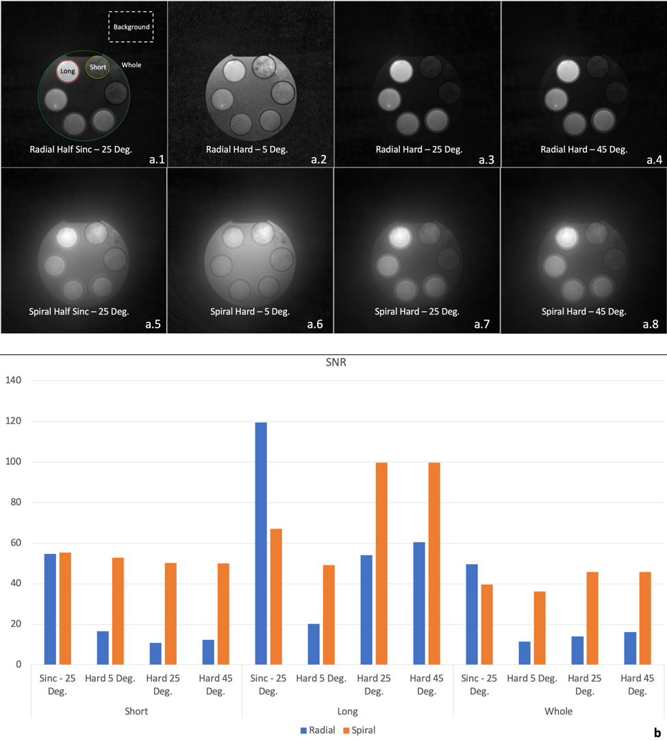
Figure 5. (a.1-8) Comparison of radial and spiral trajectories between the
half-Sinc excitation and the hard pulse excitation with different flip angles. (b)
SNR comparison.