2835
Can Accelerated SEMAC TSE Replace Conventional TSE in the Spine?1Russell H. Morgan Department of Radiology and Radiological Science, Johns Hopkins University School of Medicine, Baltimore, MD, United States
Synopsis
We investigated the exchangeability of accelerated SEMAC TSE and high-bandwidth TSE MRI in patients with spinal hardware. Two musculoskeletal radiologists found through quantitative and qualitative evaluations of 50 patients with cervical, thoracic, and lumbar spinal instrumentation that accelerated SEMAC resulted in fewer metal artifacts, better visibility of anatomical structures and abnormalities, only mildly increased blur and similar soft tissue and bone contrasts. In the spine, accelerated SEMAC TSE can be used as a replacement, rather than “add-on”, of high-bandwidth TSE.
Introduction
The prevalence of spinal fusion surgeries is steadily increasing [1]. While CT is often used to control metal artifact [2], MRI can be more powerful if metal artifacts can be sufficiently suppressed. Advanced pulse sequences using multi-spectral and multi-spatial techniques can provide substantial metal artifact reduction, but can often come with longer acquisition times, and can cause blurring and skew tissue contrast. Hence, they are often considered as “add-on”, rather than as a replacement to conventional pulse sequences. However, the integration of slice encoding for metal artifact correction (SEMAC) into a turbo spin pulse sequence in combination with parallel imaging (PI) and parameter optimization can reduce eliminate restrictions, reduce blurring, and keep acquisition times clinically feasible. Therefore, we tested the hypothesis that optimized PI-SEMAC TSE can replace high-bandwidth TSE in the spine, thereby affording improved metal artifacts reduction of spinal hardware.Methods
Following prospective IRB approval and consent, we included 50 patients (56 (18-81) years-of-age) with metallic instrumentation of the cervical, thoracic, or lumbar spine between 01/2017 and 08/2019. The MRI protocol including pairs of T1- and T2-weighted sagittal high-bandwidth (acquisition time: 4:19 min and 3:50 min, respectively) and PI-SEMAC (acquisition time: 6:39 min and 6:49 min, respectively) pulse sequences with otherwise identical sequence parameters. Following separation, blinding, and randomization, two musculoskeletal radiologists evaluated the datasets for image quality, metal artifact reduction, visibility of periprosthetic structures and abnormalities, and signal-to-noise (SNR) and contrast-to-noise (CNR) ratios of anatomical structures. Comparative and interchangeability statistics were applied. P-values < 0.05 were considered statistically significant.Results
PI-SEMAC images demonstrated significantly fewer metal artifacts compared with the high-bandwidth MRI (p<0.001). PI-SEMAC improved the visibility of anatomical structures and abnormalities and introduced slightly more blur, which were non-significant. SNR and CNR were similar for high-bandwidth and PI-SEMAC images. PI-SEMAC could substitute high-bandwidth technique, but not vice-versa.Conclusion
PI-SEMAC MRI results in significantly fewer metal artifacts of spinal hardware, better visibility of anatomical structures and abnormalities, only mildly increased blur and similar soft tissue and bone contrasts than high-bandwidth MRI. T1- and T2-weighted PI-SEMAC sequences can be used instead of high-bandwidth sequences, rather than as more time-consuming add-on sequences.Acknowledgements
No acknowledgement found.References
1. Park C, Lee E, Yeo Y, Kang Y, Lee JW, Ahn JM, Kang HS. Spine MR images in patients with pedicle screw fixation: Comparison of conventional and SEMAC-VAT sequences at 1.5 T. Magn Reson Imaging. 2018 Dec; 54:63-70.
2. Ghodasara N, Yi PH, Clark K, Fishman EK, Farshad M, Fritz J. Postoperative Spinal CT: What the Radiologist Needs to Know. Radiographics. 2019 Oct; 39(6):1840-1861.
Figures
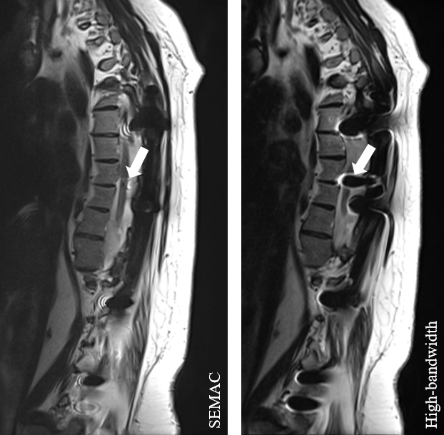
PI-SEMAC causes fewer metal artifacts (arrows) and improves visibility of anatomical structures and abnormalities compared with high-bandwidth MRI