2833
Evaluation of Knee Osteoarthritis (OA) Using 3D Ultrashort-Echo-Time Cones Magnetization Transfer (3D UTE-Cones-MT) Imaging1The third affilated hospital of Southern Medical Univerisity, Guangzhou, China, 2Department of Radiology, University of California, San Diego, CA, United States, 3Radiology Service, VA San Diego Healthcare System, San Diego, CA, United States
Synopsis
Magnetization transfer (MT) imaging has been used for indirect assessment of macromolecules in biological tissues. In this study, we explore the value of the 3D UTE-Cones-MT sequence for volumetric quantification of magnetization transfer ratio (MTR) and macromolecular proton fraction(f) in menisci of healthy volunteers and patients with different degrees of OA. The primary results demonstrate that this method can detect compositional changes in meniscus and can be used to differentiate healthy subjects from patients with mild or advanced OA.
Introduction
Because the menisci play a critical role in the long-term health of the knee joint, objective assessment of meniscal tissue quality and composition is of critical importance. Previous studies suggest that quantitative MRI values such as T1rho and T2 are very helpful for detecting the structural alterations in meniscal tissues of patients with osteoarthritis (OA) (1). However, both T1rho and T2 are difficult to detect in early meniscus degeneration, making it difficult to discriminate healthy subjects from patients with mild OA (2,3). We aimed to evaluate the 3D ultrashort echo time Cones magnetization transfer (3D UTE-Cones-MT) sequence in detecting structural alterations in meniscal tissue of healthy subjects and patients with different degrees of OA.Subjects and Methods
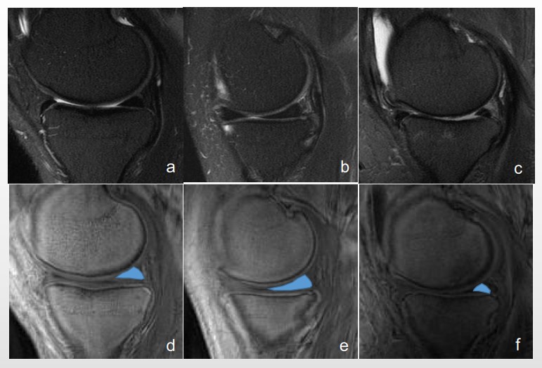
This study was approved by the Institutional Review Board. A total of 48 individuals, including healthy (n=17; 20-49 years; 7 males), mild OA (n=19; 37-86 years; 10 males), and advanced OA (n=12; 52-88 years; 4 males) subjects were recruited for this study. Clinical morphological and quantitative 3D UTE-Cones-MT images were acquired on a clinical 3T GE MR750 scanner (4,5). Morphological assessment was performed using meniscal Whole-Organ Magnetic Resonance Imaging Score (WORMS) (1-3), as shown in Figure 1. Magnetization transfer ratio (MTR) as well as two-pool MT modeling of macromolecular proton fraction (f) were calculated for medial and lateral menisci. Correlations of f and MTR with age and WORMS scores of the three groups were calculated.Results
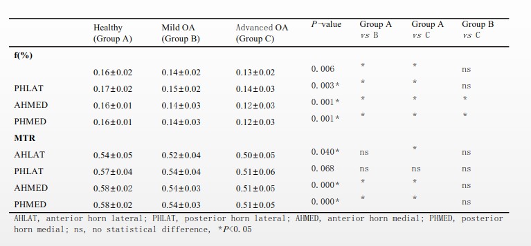
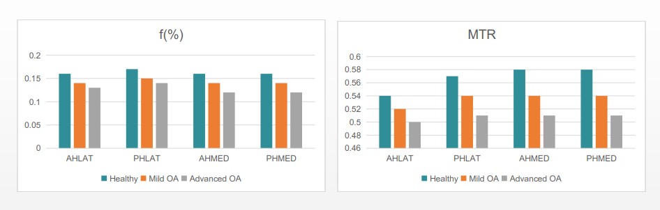
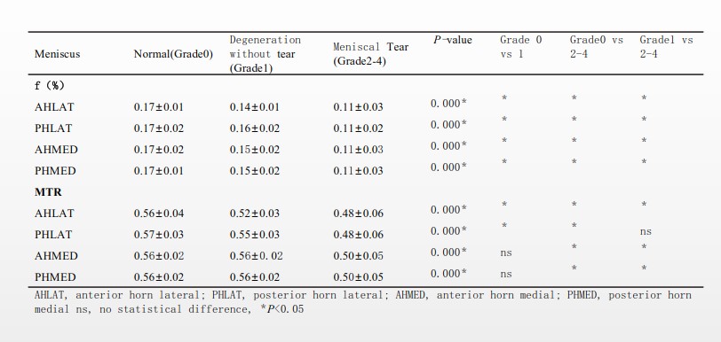
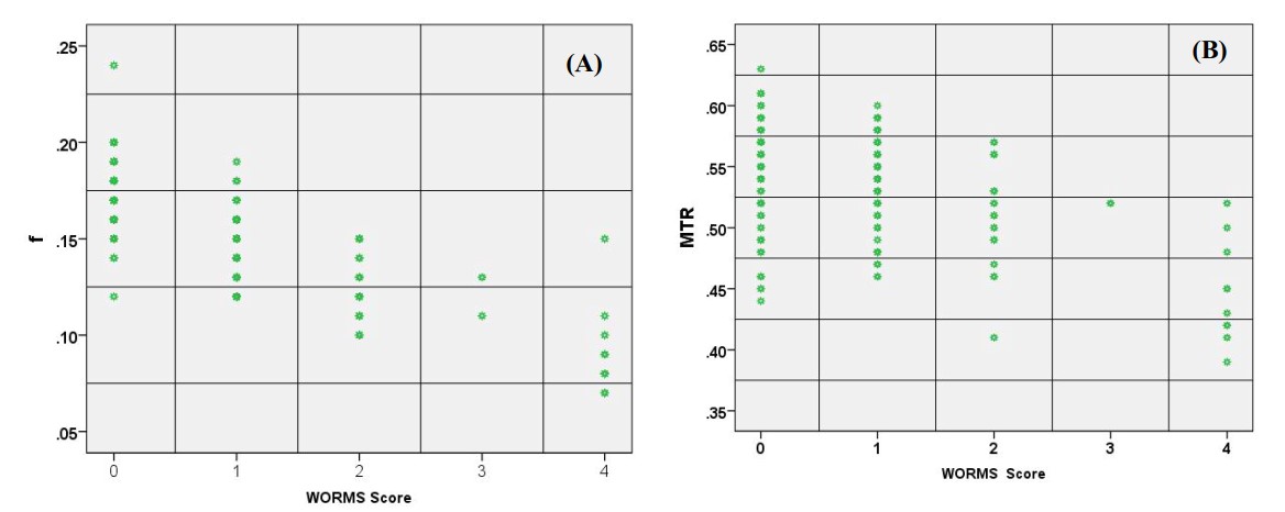
Figure 1 shows representative clinical and MT images of a healthy volunteer, a patient with mild OA, and a patient with advanced OA. The healthy meniscus showed the highest f and MTR, while the advanced OA meniscus showed the lowest f and MTR. Decreased f and MTR were observed in mild or advanced OA, as shown in Table 1 and Figure 2. Meanwhile, decreased f and MTR were observed in meniscal tear (Grade 2-4) or without tear (Grade 1) compared with normal meniscus (Grade 0), as shown in Table 2. Significant correlations were observed between f and WORMS score, as well as between MTR and WORMS scores, with r values of -0.764 and -0.323, respectively, as shown in Figure 3. P-values were less than 0.05 for both f and MTR.Discussion and Conclusion
The 3D UTE-Cones-MT measures of f and MTR correlated with meniscal WORMS grades, suggesting that UTE-Cones-MT biomarkers can detect compositional changes in meniscus and can be used to differentiate healthy subjects from patients with mild or advanced OA.Acknowledgements
The authors acknowledge grant support from NIH (1R21AR073496, R01AR068987) and VA Clinical Science and Rehabilitation R&D (I01CX001388 and I01RX002604).References
1. Meniscal Measurements of T1ρ and T2 at MR Imaging in Healthy Subjects and Patients with Osteoarthritis. Radiology: Volume 249: Number 2-November 2008.
2. Cartilage and Meniscus Assessment Using T1rho and T2 Measurements in Healthy Subjects and Patients with Osteoarthritis. Osteoarthritis Cartilage. 2010 November ; 18(11): 1408–1416.
3. Assessment of meniscus with adiabatic T1ρ and T2ρ relaxation time in asymptomatic subjects and patients with mild osteoarthritis: a feasibility study. Osteoarthritis and Cartilage 26 (2018) 580-587.
4. Ultra-high field magnetic resonance imaging parameter mapping in the posterior horn of ex vivo human menisci. Osteoarthritis Cartilage. 2018.12.
5. Ultrashort echo time magnetization transfer (UTE‐MT) imaging and modeling: magic angle independent biomarkers of tissue properties. NMR Biomed. 2016; 1–7.
Figures