2815
MRI of the knee during weight-bearing standing postures involving positions of joint extension and flexion1Medical Imaging, The First Affiliated Hospital of Nanjing Medical University, Nanjing, China, 2Jiangsu LiCi Medical Device Co. Ltd, LianYunGang, China, 3ITEE, The University of Queensland, Brisbane, Australia, 4The Australian e-Health Research Centre, CSIRO, Brisbane, Australia, 5School of Human Movement & Nutrition Science, The University of Queensland, Brisbane, Australia
Synopsis
Standing weight-bearing MR images of the knee joint in full extension and at various flexion angles (~15°, 30°) were acquired using 2D and 3D sequences on a newly developed open 0.25T extremity MRI system. 3D FLASH images were used to calculate measures of patella positioning used in previous clinical investigations as well as to produce MRI-based 3D bone models. The use of MRI and 3D modeling for topographical analyses of the bones and soft-tissues of the knee joint have the potential to provide more detailed assessment of pathoanatomical conditions involving structures such as the patella tendon and the menisci.
Introduction
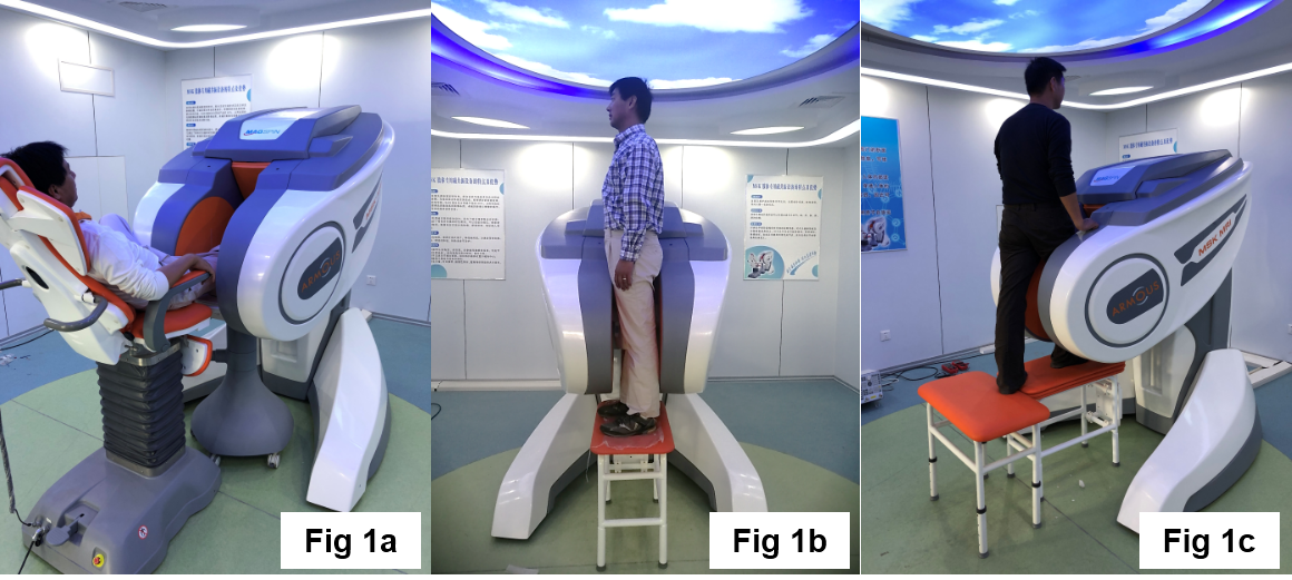
MR imaging of the knee joint under “weight-bearing” conditions has been suggested as a useful clinical approach for investigating the “naturally” loaded topographical relations between tissues and to provide diagnostic analyses not readily available using conventional recumbent positioning MRI systems [1]. Further, the potential to image collagenous structures (e.g., patellar tendon, cruciate ligaments), cartilaginous elements (e.g., articular plates, menisci), muscles (e.g., quadriceps, triceps surae) and bone units (e.g., patella, distal femur) in various degrees of knee flexion during weight-bearing MRI also offers insight into tissue topography not accessible with standard unweighted imaging. In addition, the generation of 3D models of the bones and soft-tissues of the knee joint during weight-bearing standing postures is possible based on the volumetric imaging capacities of various MRI sequences. In the current study, a 0.25T upright open extremity MRI system was used to obtain images of the knee joint in both recumbent and standing (full weight-bearing) postures with the knee joint in full extension and in ~30° flexion (Fig 1) to allow visualization of the patella-patellar tendon complex and to calculate clinical measures of patella positioning used in traditional standing X-ray investigations.Methods
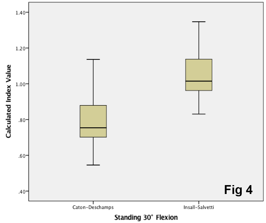
A newly-developed upright open extremity MRI system (Magspin Instrument) was used for imaging of the knee joints in 7 subjects. The dedicated extremity MRI system uses a 0.25T open permanent magnet elevated and angulated with respect to the ground. The configuration of the open magnet allows the knee joint to be imaged in conventional recumbent (resting) postures using an inclined patient chair (Fig 1a) or in weight-bearing standing postures using an elevated bench with the knee joint fully extended (Fig 1b) or with various degrees of knee flexion (Fig 1c). A two-channel orthogonal-design receive only knee coil was used to acquire 2D FSE (T1, T2 weighted), STIR and 3D FLASH images of the knee joint for the recumbent and standing postures to allow comparison of the effects of weight-bearing loads (standing postures) on tissue topography. The 3D FLASH images acquired during standing with the knee joint flexed at ~30° were post-processed with a maximum intensity projection (MIP) algorithm in SMILI [2] to generate “pseudo-Xray” images for calculation of measures (e.g., Insall-Salvati index, Caton-Deschamps index) used for clinical investigation of patella positioning. In addition, the distal femur, proximal tibia and patella were manually segmented in these FLASH images to generate 3D bone models.Results and Discussion
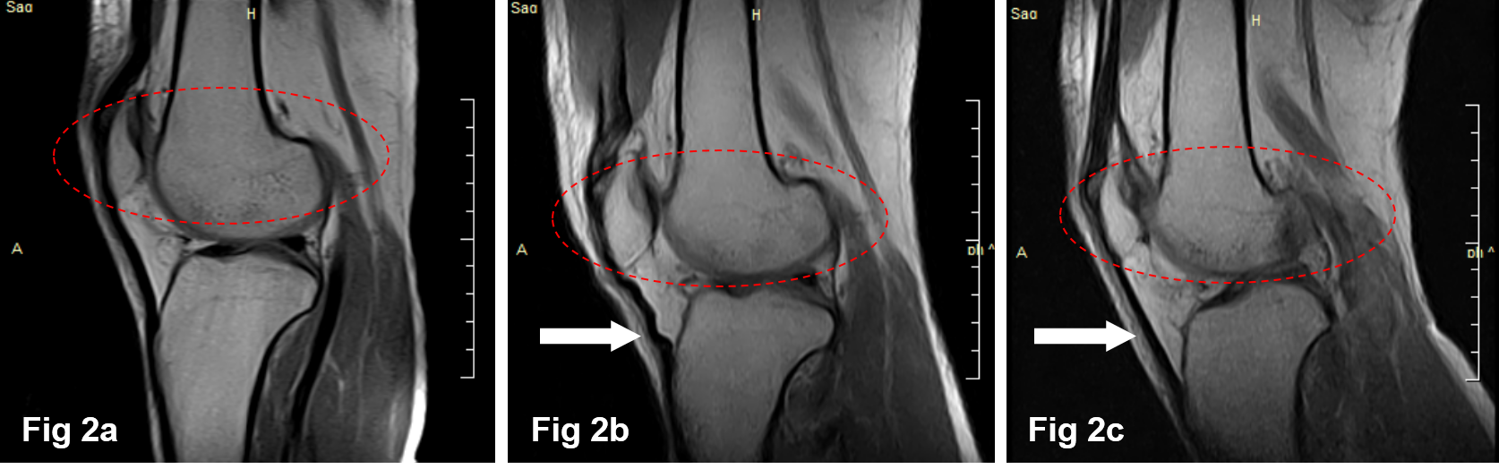
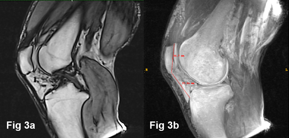
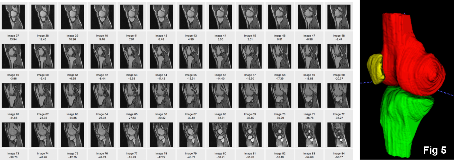
Fig 2 shows a series of T1-weighted sagittal images of the knee joint acquired in a recumbent (nonweight-bearing) position with the knee close to full extension (Fig 2a) and during standing (weight-bearing) with the knee in full extension (Fig 2b) and in ~15° of flexion (Fig 2c). The patellar tendon is well depicted in all images; in the weight-bearing standing images with knee extension there is a clear “wave” pattern of the relaxed tendon whereas in the flexed knee posture the tendon is tensioned and is seen as a straight (taut) dark band (white arrows). The inferior positioning of the patella relative to the femoral surface (red dashed oval) is also evident in the standing weight-bearing postures. Figure 3 shows an example of a single sagittal slice from the 3D FLASH sequence (Fig 3a) from which a “pseudo” 2D Xray image was generated using the MIP algorithm for calculation of either the Caton-Deschmaps index or Insall-Salvati index (shown in Fig 3b) to evaluate the relative position (height) of the patella with ~30° of knee flexion during weight-bearing standing. The Caton-Deschmaps index and Insall-Salvati index data are shown in Fig 4. Fig 5 shows an illustrative example of a series of sagittal images from the 3D FLASH sequence (in this example, images were obtained from weight-bearing standing with the knee in full extension) which were used to generate 3D bone models of the distal femur, proximal tibia and patella for qualitative topographical assessments of these bones with the knee fully extended or flexed to ~30°.Conclusion
Standing weight-bearing MR images of the knee joint in full extension and at various flexion angles (15°, 30°) were successfully acquired using a variety of 2D and 3D sequences on a newly developed open 0.25T extremity MRI system. Qualitative assessments of the patella tendon showed differences in the “waviness” of this structure in weight-bearing standing with the knee fully extended compared with ~15°-30° of flexion. The 3D FLASH sequence was used to generate a “pseudo” X-ray image to calculate measures used in previous clinical investigations of patella positioning as well as to produce MRI-based 3D bone models. In future MRI studies, the use of 3D modeling for topographical analyses of the bones and soft-tissues of the knee joint have the potential to provide more detailed assessment of pathoanatomical conditions involving structures such as the patella tendon and the menisci.Acknowledgements
No acknowledgement found.References
1. Federico B, Antonio B, Francesco A, et al. Weight-bearing MRI of the knee: a review of advantages and limits. Acta Biomed 2018; Vol. 89, Supplement 1: 78-88.
2. Chandra S, Dowling J, Engstrom C, et al. A lightweight rapid application development framework for biomedical image analysis. Computer methods and programs in biomedicine 2018; 164: 193-205.
Figures