2814
Dynamic multinuclear measurements in the calf with a dedicated RF coil while taking into account the nuclear Overhauser enhancement effect.1NMR Laboratory, Institute of Myology, Paris, France, 2NMR Laboratory, CEA/DRF/IBFJ/MIRCen, Paris, France, 3Siemens Healthcare SAS, Saint-Denis, France
Synopsis
Interleaving multinuclear NMR measurements in the muscle during a transient state enriches the value of the exam by generating multiple data sets simultaneously. Here we show the results of our multinuclear interleaved approach using a 1H/31P dual-tune RF coil dedicated for calf experiments during an exercise and an ischemia-hyperemia paradigm. The nuclear Overhauser enhancement effect, which is dependent on the sequence and RF design, is characterized and its impact on rephosphorylation rate estimation and the [Pi]/[PCr] ratio is evaluated. The proposed methodology constitutes a powerful tool to evaluate mitochondrial function and metabolic efficiency in vivo.
INTRODUCTION
NMR allows investigating a plethora of physiological and biochemical variables in vivo during a dynamic paradigm. Interleaving multinuclear NMR measurements during a transient state enriches the value of the exam by generating multiple data sets and, by simultaneously acquiring them, multifactorial variables can be investigated1,2 without introducing experimental variability concerns3 from repeating the experiment multiple times as with traditional sequences. We show the results of a new calf setup during dynamic experiments using a novel, dedicated dual-tune RF coil where concomitant T2* maps and work measures have been added with respect to previous work done at 3T4. The nuclear Overhauser enhancement (nOe) effet on 31P metabolites, which depends on the specific 1H RF pulses and sequence timings used, is characterized at rest for the current setup. Potential discrepancies of dynamically measured variables are investigated by performing two exercise paradigms, one using our 1H/31P multinuclear interleaved sequence and the other one using only its 31P FID module.METHODS
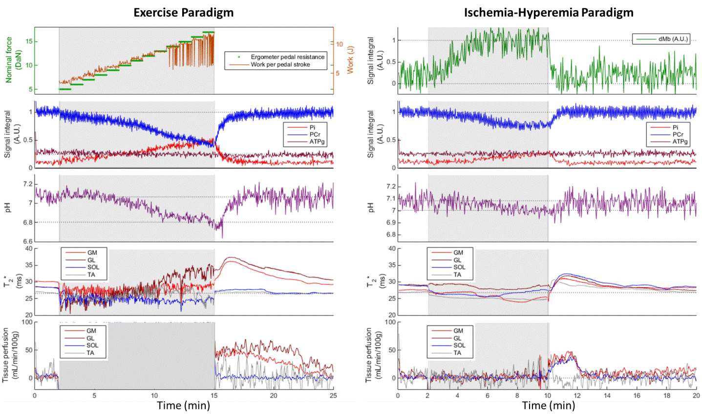
Experimental Setup9 healthy subjects (34 ± 16 y.o., 6 females) participated in this study. Experiments were done on a 3-T/60-cm bore Siemens Magnetom Prisma MR system. The RF coil used (RAPID Biomedical) combined a 1H birdcage transmitter (20-cm inner diameter, 30-cm resonator length), a 1H 18Rx semi-flexible phased-array receiver and a 31P 1Tx/3Rx semi-cylindrical transceiver. The calf of the subject was placed facing the 31P coil and at the distal end of the 1H coils. Each subject performed in a single visit a leg ischemia and two identical exercise paradigms, using an amagnetic ergometer, as detailed in fig.1A.
Interleaved NMR
A non-localized 31P MRS spectrum and 32 non-localized 1H MRS spectra resonating at the deoxygenated myoglobin frequency(dMb) were acquired during the evolution time (820 ms) of a pulsed-ASL (SATIR5) sequence generating a perfusion-weighted 2D radially-encoded image followed by a multi-echo 2D GRE radial acquisition every 2.5 seconds (fig.1B). The interleaved sequence was used during the first exercise and the ischemia-hyperemia paradigm. 14 minutes after the first exercise, the nOe effect of each 1H RF pulse was measured for 1 minute by turning off the other 1H pulses. During the second exercise paradigm, only the 31P NMRS module and magnetic field gradients were activated.
Data Analysis
Raw data was reconstructed and quantified using in-house Matlab routines. No motion correction was applied. Dynamic 31P spectra were phased automatically and visually checked. The 31P spectra used to measure the nOe effect were manually phased for zero- and first-order phase corrections. dMb spectra were phased using a water reference and were averaged before baseline correction. Images were reconstructed using a non-uniform FFT algorithm6. Perfusion values were calculated from every alternating pair of images as described previously5. T2* values and the myoglobin resaturation rate(τMb) were calculated using an exponential decay. The rephosphorylation rate of PCr (τPCr) was estimated with an exponential function during exercise recovery.
RESULTS AND DISCUSSION
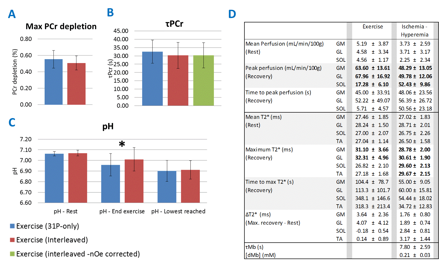
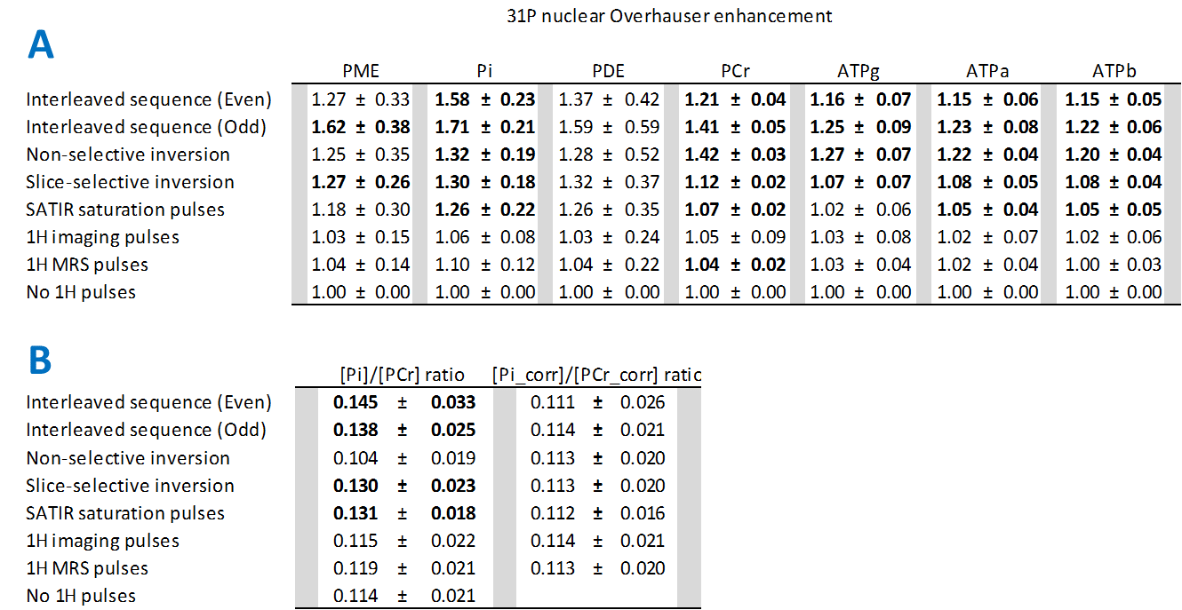
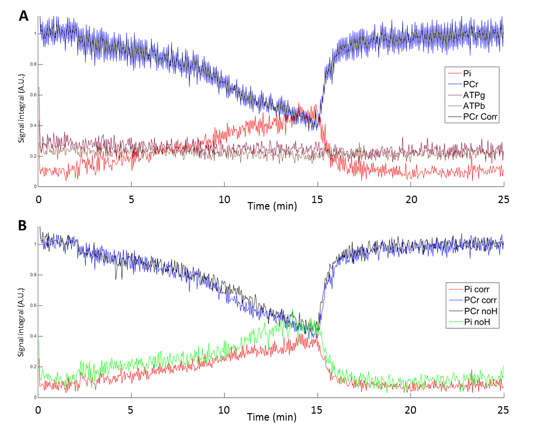
Multi-parametric time courses from the same subject are shown for the exercise and ischemia-hyperemia paradigms (fig.2). The extracted parameters are summarized in fig.3 and the metabolite-specific nOe effects generated by each RF pulse are shown in fig.4A.The two exercises showed no significant difference of PCr depletion (fig.3A), initial pH or lowest pH reached (occurring shortly after the end of exercise). A lower pH at the end of exercise was found for the 31P-only measurements (fig.3C). No differences were found (P=0.273) for τPCr between the 31P-only and interleaved data (32.57±7.0 s and 30.27±7.8 s, respectively). For each subject, applying the “interleaved seq. even” and “interleaved seq. odd” nOe corrections of PCr to the even and odd repetitions of the [PCr] interleaved data resulted in a τPCr-nOe corrected value (fig.3B) which is not statistically different than the uncorrected τPCr (P=0.28). Dynamic time courses of the two exercises from one subject, before and after nOe corrections, are shown in fig.5.
The impact of nOe was also evaluated on the [Pi]/[PCr] ratio at rest (fig.4A). Significant alterations were induced by the SATIR slice-selective pulses and by the standard interleaved sequence during both odd and even repetitions (fig.4B, left column). Correcting the nOe effect on [Pi] and [PCr] for each subject using the values shown in fig.4A restores the [Pi]/[PCr] ratio to values close to the 31P-only sequence(fig.4B, right column).
The measured absolute dMb concentration, blood tissue perfusion and T2* of the ischemia-hyperemia paradigm were in good agreement with other studies7,8, despite that the ischemic period used here was longer. In the exercise paradigm, these same variables showed a large increase in the gastrocnemius muscles only, as expected for a plantar flexion exercise with the leg in a straight position.
CONCLUSION
A robust experimental calf setup is presented where the potential of multinuclear interleaving is demonstrated on two dynamic paradigms. nOe effects were found to be negligible on the estimation of τPCr. The [Pi]/[PCr] ratio is nevertheless altered but a correction factor averaged from a small group of subjects tackled this issue. This setup permits to combine information from an increasingly demanding physical effort with the respective oxygen and metabolic responses and thus constitute a powerful tool to evaluate mitochondrial function9 and metabolic efficiency in vivo. Repeatability studies are currently under way.Acknowledgements
No acknowledgement found.References
1.
Toussaint JF, Kwong KK et al. J Appl Physiol. 1996; 81: 2221–2228. F(dMb)
2. Gillies RJ. NMR in Physiology and Biomedicine. Academic Press: San Diego, CA, 1994.
3. Carlier PG, Bertoldi D et al. NMR Biomed. 2006; 19:954-967.
4. Lopez Kolkovsky AL, Marty B et al. ISMRM. 2017; 89.
5. Raynaud JS, Duteil S et
al. MRM. 2001; 46 :305-311.
6. Fessler, JA & Sutton BP. IEEE Trans on Signal
Processing. 2003; 51(2):560-574.
7. Englund EK, Langham MC et al. Circ Cardiovasc Imaging. 2015; 8(4)
8. Vanderthommen M, Duteil S et al J Appl Physiol 2003, 94:1012-1024
9. Kemp GJ, Ahmad RE, et
al. Acta Physiol 2015, 213, 107-144
Figures