2811
Pre- to post-ovulatory changes in ACL T2* metrics over the course of the female menstrual cycle: A new biomarker for ACL injury risk?
Erin C Argentieri1, Tatum W Braun1, Ryan E Breighner1, Bin Q Lin2, Ellen K Casey3, Shari T Jawetz1, Alissa J Burge1, Matthew F Koff1, and Hollis G Potter1
1Radiology and Imaging, Hospital for Special Surgery, New York, NY, United States, 2Biostatistics, Hospital for Special Surgery, New York, NY, United States, 3Physiatry, Hospital for Special Surgery, New York, NY, United States
1Radiology and Imaging, Hospital for Special Surgery, New York, NY, United States, 2Biostatistics, Hospital for Special Surgery, New York, NY, United States, 3Physiatry, Hospital for Special Surgery, New York, NY, United States
Synopsis
This study evaluates changes in ACL T2* metrics over the course of the female menstrual cycle. In the pre-ovulatory phase, normally ovulating case subjects demonstrated significant shortening of T2*S and PS in comparison to visit #4 (post-ovulatory phase). Non-ovulatory control subjects displayed no significant changes over time. These findings suggest that shifts in collagen bound water occur within the ACL over the course of the menstrual cycle. Shifts in tissue water content have been associated with altered mechanical properties, and changes in ligament stiffness may alter proprioceptive sense, contribute to increases in laxity, and alter ACL-injury risk.
Introduction
Regardless of surgical or non-surgical intervention, anterior cruciate ligament (ACL) rupture is associated with the development of post-traumatic osteoarthritis (PTOA) within 5-10 years of the incident injury.1-3 As such, recent efforts have focused on identifying the risk factors for ACL-injury, with the goal of reducing its overall incidence. Previous work has identified both intrinsic and extrinsic factors that contribute to ACL-injury risk, and multiple studies have established that female ACL-injury risk is more than double that of their male counterparts.4-7 Sex based disparities in ACL-injury risk may be explained by cyclic variations in hormone levels, as previous studies have identified that female ACL-injury risk is increased along with knee joint laxity during the pre-ovulatory phase of the menstrual cycle.5,6 These findings suggest that sex hormones may directly impact the structure and biomechanical integrity of the ACL. Utilization of ultra-short echo (UTE) sequences allows for the rapid transverse relaxation times associated with short T2 species to be captured, permitting quantitative evaluation of tissue microstructure with T2* mapping.8-11 Recently, UTE-MRI and bi-component T2* analyses have been used to evaluate both the short T2* (associated with bound water) and long T2* components (associated with free water)8,9 within tissues and assess their relationship to mechanical properties.8-12 The objective of this study was to determine if significant changes in ACL T2* metrics exist over the course of a menstrual cycle. We hypothesized that normally ovulating pre-menopausal females would exhibit significant changes in ACL T2* metrics over the course of a menstrual cycle, while no significant differences would exist within anovulatory control subjects.Methods
This was an IRB approved pilot study of 15 females with no history of injury to either knee. Seven pre-menopausal females with normal menstrual cycles and no history of hormonal contraceptive use (>1 year) were included as ovulatory case subjects. Subjects within the non-ovulatory control group included 4 post-menopausal females, and 4 pre-menopausal subjects taking oral contraceptives with normal menses. All subjects participated in 1 study visit per week (same day/time each week) for a total of 4 study visits over the course of 1 month. Study visit #1 coincided with onset of menses (within 24hrs) for all pre-menopausal females. Subjects were provided with commercially available ovulation predictor kits (ClearBlue Digital Ovulation Tests [Accuracy 99%]) in order to determine date of ovulation in case subjects, and to confirm anovulatory status in control subjects. MRI Acquisition: Bilateral 3-Tesla MRI examinations were obtained on a clinical scanner (GE Healthcare) using an 8-channel phased array knee coil (Invivo). Three-dimensional, double coronal oblique UTE sequences were acquired for evaluation of T2* metrics (Voxel: 0.50x0.50x1.5mm3, TEs: 11 echoes between 0.03-25ms, TR: 166ms, RBW: ±83.3kHz, Flip-Angle: 16o). Imaging Analysis: Bi-exponential fits of SI to corresponding echo time were used to calculate ACL T2* metrics12: SI(TE) = A(-TE/T2*S) + B(-TE/T2*L)+noise, where T2*S and T2*L are respective short and long T2* components, A and B are corresponding short and long apparent proton densities, and PS is the short component fraction calculated as A/(A+B).12 Statistical Analysis: Generalized estimating equation modeling was used to cluster data points contributed from each leg of each patient, and longitudinal analyses were completed for each T2* metric (T2*S, T2*L, PS) within and between groups. Maximum likelihood estimates were used to establish parameter estimates for each study visit. Post-hoc pairwise evaluations with Bonferroni-adjustment for multiple comparisons were used to identify differences between visits.Results
Significant differences were found within and between study groups for T2*S and PS T2* metrics. At study visit #1 (pre-ovulatory phase), normally ovulating case subjects exhibited significantly decreased T2*S and PS metrics compared to study visit #4 when all subjects were in the post-ovulatory phase (mean difference T2*S: −1.1ms; p = 0.005; mean difference PS: −1.72%; p = 0.02). Anovulatory control subjects exhibited no such significant changes over time for any T2* metric. (Figure 1)Discussion
ACLs of normally ovulating, pre-menopausal case subjects displayed significant changes in T2* metrics over the course of a menstrual cycle, while anovulatory control subjects displayed no such differences. Specifically, significant differences were exhibited between study visit #1, when all case subjects were in the pre-ovulatory phase, and study visit #4 when all case subjects were in the post-ovulatory phase. These findings suggest that shifts in ACL tissue water content occur over the course of the menstrual cycle; the relative shortening of T2* metrics within the pre-ovulatory phase may be indicative of an increase in collagen bound water (T2*S).8, 9 Previous studies have demonstrated that shifts in tissue water content are associated with altered mechanical properties, and decreased T2* has been demonstrated in preclinical models of cyclic loading prior to gross disruption of collagen fibrils.10,11 Subsequent changes in ligament stiffness over the course of the menstrual cycle may alter proprioceptive sense and contribute to the observed increases in both knee laxity and ACL-injury risk during the pre-ovulatory phase of the menstrual cycle.Conclusion
This is the first study to evaluate changes in ACL T2* metrics throughout the menstrual cycle. These data suggest that significant differences exist between pre- and post-ovulatory phases of the menstrual cycle, and may be indicative of a new imaging biomarker for ACL-injury risk.Acknowledgements
HSS has an institutional research agreement with GE Healthcare. The authors would like to thank Kelly Zochowski, Erica Hooper, and Sade Clark as well as the entire HSS MRI staff and technologists for their assistance with this studyReferences
(1) Oiestad 2010; (2) Potter 2012; (3) Lohmander 2007; (4) Beynnon 2014; (5) Beynnon 2006; (6) Shultz 2005; (7) Beynnon 2015; (8) Pauli 2012; (9) Diaz 2012; (10) Jerban 2017; (11) Koff 2014; (12) Juras 2013Figures
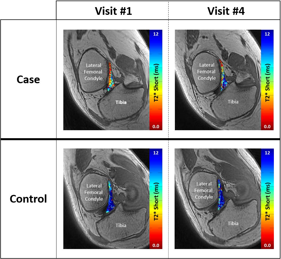
Figure 1: Example of changes in ACL T2*S
within ovulating
case
subjects (top) and anovulatory control subjects (bottom) from study
visit #1 (pre-ovulatory) to study visit #4 (cases post-ovulatory). Significant
changes were found within case subjects only.