2798
AcidoCEST-UTE MRI is a Reproducible Method to Measure Cartilage and Meniscus pH1Radiology, University of California San Diego, San Diego, CA, United States, 2Radiology Service, VA San Diego Healthcare System, San Diego, CA, United States, 3Orthopedic Surgery Service, VA San Diego Healthcare System, San Diego, CA, United States
Synopsis
The acidification of musculoskeletal tissues is being investigated as a way to localize pain in osteoarthritis. AcidoCEST-UTE MRI can measure pH in tissues such as cartilage and meniscus, though in vivo validation is difficult. Thus, it is imperative that the acidoCEST-UTE sequence be reproducible and verifiable ex vivo, where pH measurements can be confirmed with a pH electrode. This study tested the reproducibility of the acidoCEST-UTE sequence in three ex vivo phantoms, confirming the pH sensitivity of the technique and demonstrating the consistency of the method and results.
Introduction
Osteoarthritis (OA) has a substantial impact on affected individuals and on the healthcare system as a whole. While diagnostic imaging of OA is widely practiced, structural degradation is often poorly correlated with pain reported by individuals. Acidification is a major component in the progression of pain,1,2 which could help localize and potentially be corrected to treat patient discomfort.Non-invasive, pH-sensitive imaging is possible with chemical exchange saturation transfer (acidoCEST) MRI. Recently, Ma et al.3 modified acidoCEST MRI for use in short T2 musculoskeletal tissues such as cartilage and meniscus by adding a 3D ultrashort echo time (UTE) readout to the acidoCEST sequence. This method has been used successfully to measure pH in ex vivo tissue phantoms as well as in vivo.4 Due to the difficulty of validating pH in vivo, we have conducted repeated measurements in ex vivo tissue to test the stability and accuracy of acidoCEST-UTE MRI.
The purpose of this study was to verify the reproducibility of the acidoCEST-UTE sequences in musculoskeletal tissue for pH measurement.
Methods
Phantom Preparation:Thirty-three syringes were filled with either iohexol or iopamidol at 200 mM concentration and PBS at 10 mM and pH-adjusted in a range from 6.2 to 7.8. Coins of pure cartilage and meniscus were harvested from grossly normal tissues from ten donors (nine male, one female; age range 25-80), soaked for 24 hours in the same buffered contrast agent solutions as described above, and included in the syringes. Syringes were grouped into three sets and each set was placed in an agar-filled container (henceforth referred to as Phantom 1-3) and scanned parallel to B0 at isocenter. All three Phantoms were imaged with the same sequence on three different days.
MRI and Analysis:
Liquid and tissue phantoms were imaged on the same 3T clinical scanner (MR750, GE Healthcare, Milwaukee, WI) using an 8-channel transmit/receive knee coil.
The 3D acidoCEST-UTE-Cones sequence3 parameters are shown as follows: TR=62ms, TE=0.032ms, Nsp=5, spoke interval τ=5 ms, FA=5°, BW=166 kHz, FOV=12×12×11.2cm3, matrix=160×160×28. CEST contrast was created with a Fermi pulse: duration=32ms, bandwidth=40Hz, two average B1s=5.4/1.1µT, 61 frequency offsets from -1200 to 1200 Hz in 40 Hz increments, with 1min 10s scan time per frequency. Steady-state before acquisition was achieved using an 8s dummy scan.
AcidoCEST MRI data was analyzed using ratio of power mismatch (RPM) based on distinct RF-powers (0.54 and 1.10 µT)5 at the 4.3 ppm peak for iohexol or the 4.2 ppm peak for iopamidol. For individual phantoms, correlations were preformed between pH and RPM for each contrast and tissue. In addition, pH was modeled as a function of RPM for the three phantoms combined, goodness of fit was measured, and a 95% confidence ellipse plot was generated.
Results
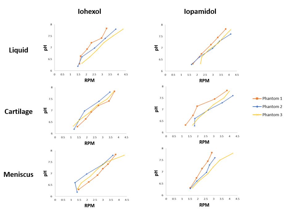
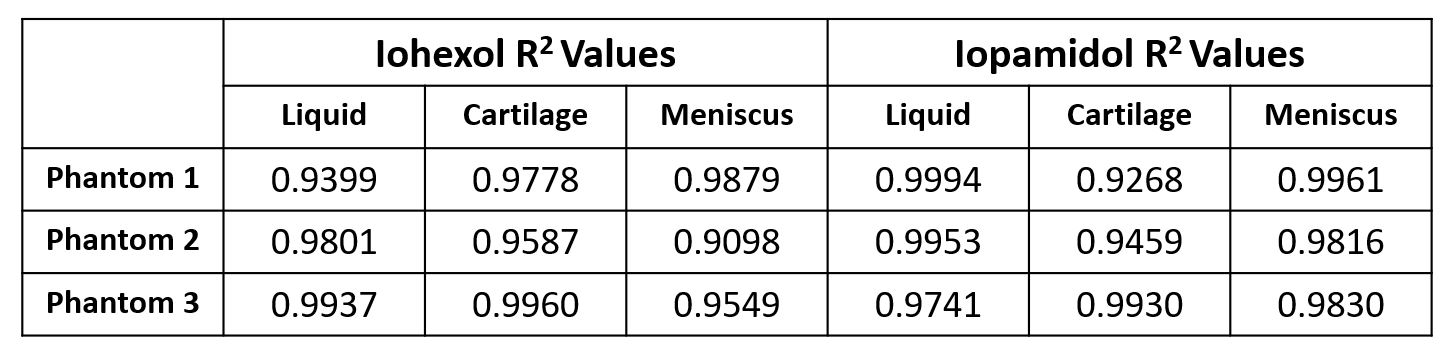
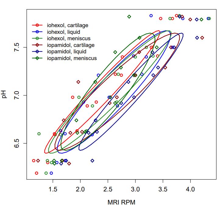
The relationship between RPM and pH shows good agreement across all three phantoms for both iohexol and iopamidol (Figure 1). R2 values for each phantom are given in Figure 2. For the three phantoms combined, excellent goodness of fit was demonstrated (iohexol R2 range 0.854-0.946, iopamidol R2 range 0.792-0.934), indicating strong reproducibility. The 95% confidence ellipses are shown in Figure 3. For both iohexol and iopamidol, confidence intervals overlap between liquid, cartilage, and meniscus, indicating no statistically significant difference.Discussion and Conclusions
In vivo pH measurements are problematic to accurately verify. Imaging methods such as acidoCEST MRI must therefore first be validated ex vivo to demonstrate reproducibility and accuracy of their measurements. In this study, we show the repeatability of acidoCEST-UTE MRI across three individually made phantoms containing cartilage, meniscus, and iohexol or iopamidol.Acknowledgements
The authors gratefully acknowledge grant support from the VA Rehabilitation R&D Service (I01RX002604), VA Clinical Science R&D Service (I01CX001388) and NIH (R21AR073496, R01AR062581, R01AR075825, and 1R01AR068987).References
1. Abdelhamid RE and Sluka KA. ASICs mediate pain and inflammation in musculoskeletal diseases. Physiology. 2015 Nov;30(6): 449-59.
2. Reeh PW and Steen KH. Tissue acidosis in nociception and pain. Prog Brain Res. 1996;113: 143-51.
3. Ma Y-J, High RA, Tang Q, Wan L, Jerban S, Du J, Chang EY. AcidoCEST-UTE MRI for the assessment of extracellular pH of joint tissues at 3T. Invest Radiol. 2019;54(9): 565-571.
4. High RA, Ji Y, Ma Y-J, Tang Q, Murphy ME, Du J, Chang EY. In vivo assessment of extracellular pH of joint tissues using acidoCEST-UTE MRI. Quant Imaging Med Surg. 2019;9(10): 1664-73.
5. Wu R, Longo DL, Aime S, Sun PZ. Quantitative description of radiofrequency (RF) power-based ratiometric chemical exchange saturation transfer (CEST) pH imaging. NMR Biomed. 2015 May;28(5): 555-65.
Figures