2796
Evaluation of the 3D VISTA sequence with compressed sensing acceleration for imaging of the ankle: a Preliminary study1Departments of Radiology, Union Hospital, Tongji Medical College, Huazhong University of Science and Technology, Wuhan, China, 2Philips Healthcare, Beijing, China
Synopsis
The three-dimensional (3D) high resolution volume isotropic FSE sequence of ankle magnetic resonance imaging has the capabilities of acquiring thin-sliced sections and performing MPR, at the cost of long scan time. Several recent studies showed that the compressed sensing(CS) helps to reduce scan time, which is especially beneficial in 3D imaging. The aim of this study was to investigate the feasibility of 3D VISTA imaging with CS in ankle joint imaging, in comparison to 3D-VISTA with parallel imaging (PI ). The results showed that the acquisition time of 3D-VISTA MRI was reduced with CS while the image quality was retained.
INTRODUCTION
Sprain of the ankle joint is one of the most common sports injuries. The ankle anatomy is complex, and the ligament tendons are distorted, making it difficult to display in the same slices on the 2D images. The three-dimensional (3D) volume isotropic turbo spin echo acquisition (VISTA) sequence of ankle magnetic resonance imaging(MRI) has the capabilities of acquiring thin-sliced sections and performing MPR without any loss of spatial resolution, which would be widely used for preoperative evaluation of surgery and 3D printing[1, 2]. However, The long scan time for 3D-VISTA imaging relays a major impediment to use isotropic high spatial resolution 3D-VISTA sequences, as it could cause motion artifacts and patient discomfort [3]. In several recent studies, the compressed sensing(CS) technique has been applied to the 3D sequence of MRI, which helped in the scan time reduction while showing minimal effect on image quality[4, 5].As far as we know, there were few studies focus on 3D-VISTA MRI with compressed sensing for the ankle joint. This study was to investigate the feasibility of 3D VISTA imaging with CS compared to 3D-VISTA imaging with parallel imaging (PI ) in evaluating ankle joint.METHOD AND MATERIALS
Institutional review board approval and informed consent were obtained. Eight volunteers with 16 ankles and two patients with ankle injury underwent 3T MRI (Ingenia CX; Philips Healthcare, Best, the Netherlands) of the ankle with eight-channel ankle joint coil, including acquisition of image sets of 2D-FSE sequences, and 3D-VISTA sequences with CS factor 6 and PI factor 3 (2x1.5) . All scans were evaluated by 2 musculoskeletal radiologists concerning image quality of the 3D-VISTA-CS and 3D-VISTA-PI sequence, and scored the inter-sequence agreement using a four-point scale. Quantitative image similarity and objective image quality were evaluated by calculating structural similarity index (SSIM). To compare the SNR and CNR between the two sequences, paired t-test was used. The Wilcoxon signed-rank test was used for comparison of the image quality scores between the two sequences. A P-value of less than 0.05 was considered statistically significant.RESULTS
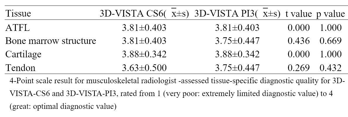
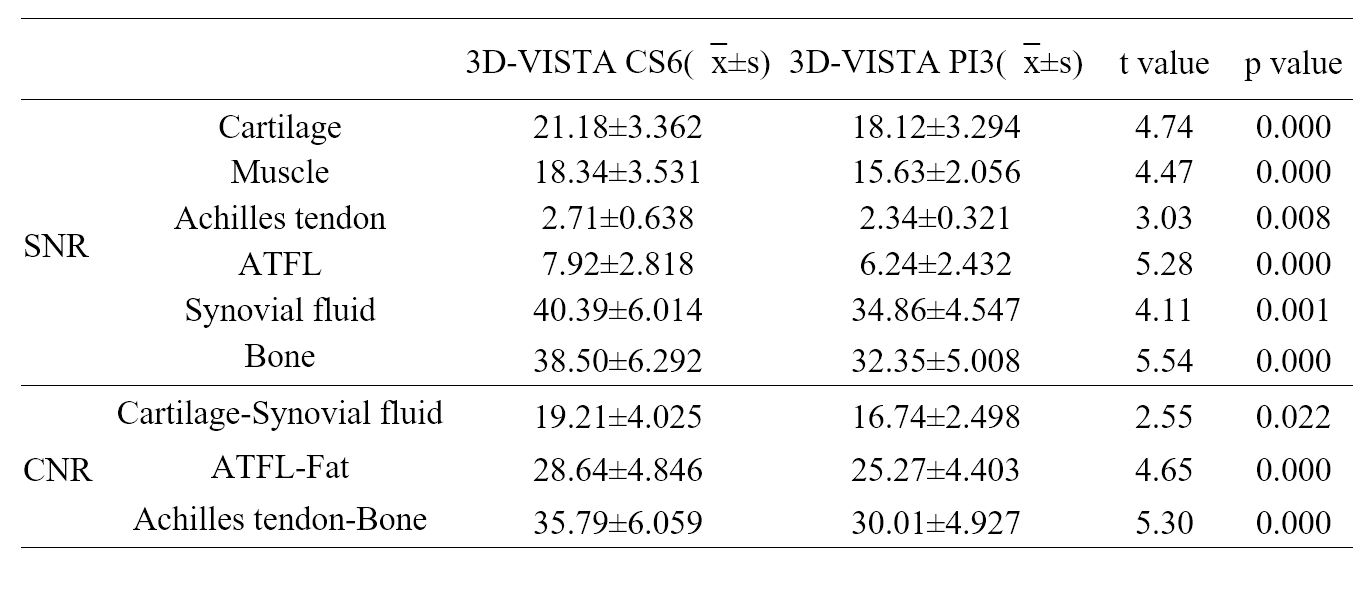
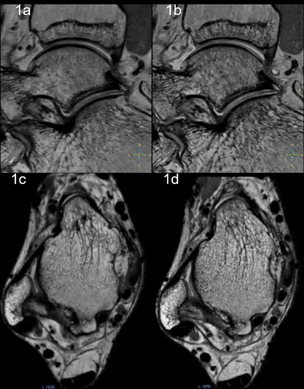
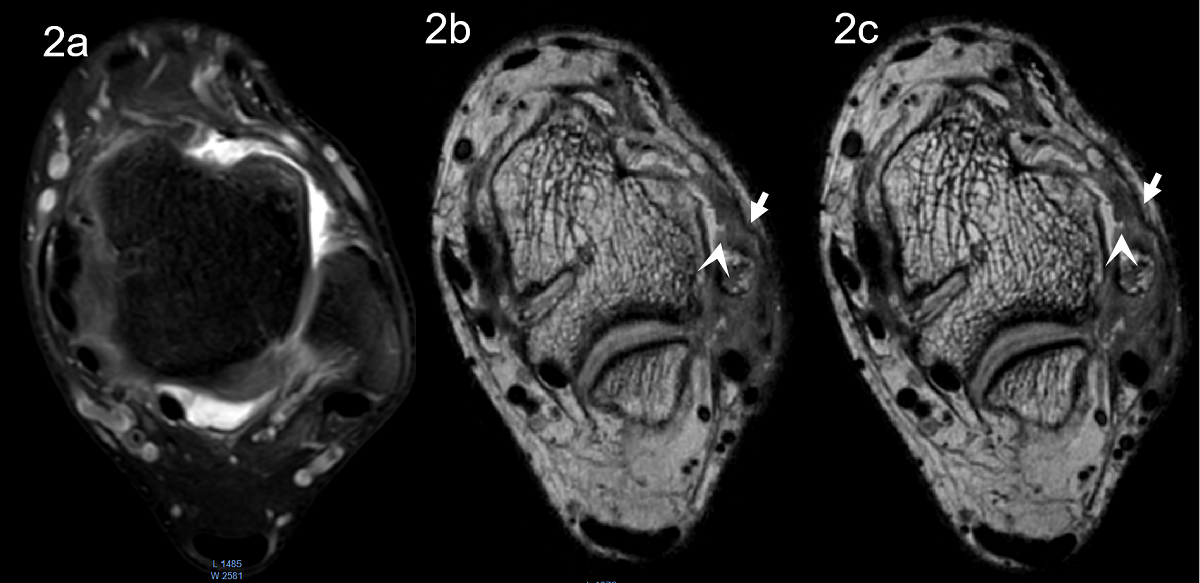
The acquisition time of 3D-VISTA MRI was reduced with CS (11 min 47 s vs. 5 min 50 s).For perceived tissue-specific diagnostic quality as assessed by two musculoskeletal radiologists, there was no significant difference between 3D-VISTA with CS6 and 3D-VISTA with PI3 (Figue 1) on evaluation of anterior talofibular ligament(ATFL)(p=1.00), bone marrow structure (P=0.669), cartiage(P=1.00) and tendon(P=0.432, Table 1). For calculated tissue-specific signal characteristics, the SNRs of 3D-VISTA with CS were slightly higher than that of 3D-VISTA with PI in tissues of cartilage, muscle, achilles tendon, ATFL, synovial fluid and bone. All CNRs of bone marrow–achilles tendon, cartilage- synovial fluid and ATFL-surrounding fat were measured slightly higher in images obtained with CS than in those obtained with PI with significant statistical difference(Figure 2). (P<0.05,Tables 2). The structural similarity index (mean, 0.995; range, 0.991-0.997) between the 3D-VISTA sequences with CS and 3D-VISTA with PI was acceptable.DISCUSSION AND CONCLUSION
With the same imaging parameters, 3D-VISTA-CS has halved the scan time of 3D-VISTA-PI, while the subjective scores of the image quality on each fine-structure were retained. Quantitative comparisons between the sequences suggested higher SNR and CNR of the images from 3D-VISTA-CS scan. However, the general signal intensities were consistent between the two sequences, implying the higher SNR from 3D-VISTA-CS scan was likely due to the reduced noise through the iterative reconstruction[6]. Images acquired with both sequences achieved good similarity as measured using SSIM, implying the well preservation of the fine-structure in the 3D-VISTA-CS scan, despite the reduced k-space samplings. In conclusion, implementing compressed sensing with 3D-VISTA scan helps to dramatically reduce the imaging time in ankle and the potential image quality degradation, if any, is neglectable. A limit of the study is the relatively small sample size and the use of healthy volunteers.Acknowledgements
Many thanks to Department of Radiology, Union Hospital, TongjiMedical College, Huazhong University of Science and Technology. Many thanks to MR Collaborations, Philips Healthcare, Beijin , China.
References
1. Jung H-G, Kim N-R, Kim T-H, Eom J-S, Lee D-O: Magnetic Resonance Imaging and Stress Radiography in Chronic Lateral Ankle Instability. Foot & Ankle International 2017, 38(6):621-626.
2. Park HJ, Lee SY, Park NH, Rho MH, Chung EC, Park JH, Park SJ: Three-dimensional isotropic T2-weighted fast spin-echo (VISTA) ankle MRI versus two-dimensional fast spin-echo T2-weighted sequences for the evaluation of anterior talofibular ligament injury. Clinical Radiology 2016, 71(4):349-355.
3. Henninger B, Raithel E, Kranewitter C, Steurer M, Jaschke W, Kremser C: Evaluation of an accelerated 3D SPACE sequence with compressed sensing and free-stop scan mode for imaging of the knee. European Journal of Radiology 2018, 102:74-82.
4. Lee SH, Lee YH, Song H-T, Suh J-S: Rapid acquisition of magnetic resonance imaging of the shoulder using three-dimensional fast spin echo sequence with compressed sensing. Magnetic Resonance Imaging 2017, 42:152-157.
5. Yi J, Lee YH, Hahn S, Albakheet SS, Song H-T, Suh J-S: Fast isotropic volumetric magnetic resonance imaging of the ankle: Acceleration of the three-dimensional fast spin echo sequence using compressed sensing combined with parallel imaging. European Journal of Radiology 2019, 112:52-58.
6. Lustig M, Pauly JM: SPIRiT: Iterative self-consistent parallel imaging reconstruction from arbitrary k-space. Magn Reson Med 2010, 64(2):457-471.
Figures