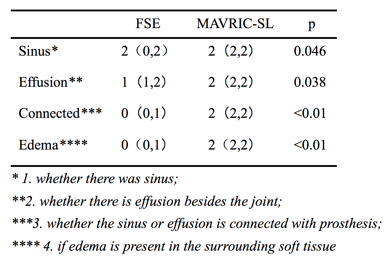
2792
Diagnostic value of MAVRIC-SL sequence in patients with suspected infection after orthopedic metal implantation1the Department of Radiology, First Affiliated Hospital of China Medical University, Shenyang,Liaoning, China, 2GE Healthcare, MR Research, Beijing, China
Synopsis
Multiacquisition with variable resonance image combination (MAVRIC-SL) sequence is a novel metal artifact reduction technique that can effectively reduce metal artifacts in patients who undergo joint replacement or fracture internal fixation. The purpose of this study was to investigate the diagnostic advantages of the MAVRIC-SL sequence MAVRIC-SL compared with conventional FSE sequence for patients with orthopedic metal implants who are suspected of having the infection.
Introduction
Prosthesis infection after orthopaedic surgery is a common and challenging complication for both patients and surgeons[1]. MRI has good soft-tissue contrast and high diagnostic value in these kinds of complications. Yet, the excessive metal implant artifacts in the conventional sequence may obscure the lesions and in turn lead to misdiagnosis in patients with suspected infection after operation. MAVRIC-SL sequence is a novel metal artifact reduction technique, which can effectively reduce metal artifacts and improve the observation of the structure around the prosthesis [2].Methods
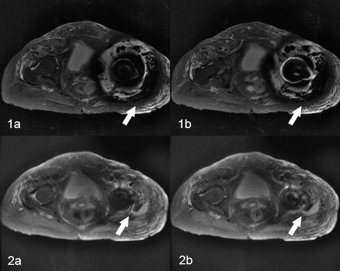
A total of 11 patients (males: 7 cases; females: 4 cases), aged 23-74 years (54.91±19.13 years), were included in this study between July, 2018 and March, 2019. All patients had orthopedic metal implants and were diagnosed with suspected postoperative infections. There was 1 case of clavicle, 1 case of humerus, 1 case of lumbar vertebrae, 5 cases of hip joint, 2 cases of knee joint and 1 case of tibia. All patients were scanned using 3.0T SIGNA PIONEER MRI system, FSE and MAVRIC-SL; T1WI, PDWI, IR were also scanned (Figure 1). Two experienced radiologists read the images and graded the conventional sequences and MAVRIC-SL. Any disagreements were resolved with consensus. The evaluation system included (1) whether there were sinuses; (2) whether there was effusion besides the joint; (3) whether the sinus or effusion was connected with prosthesis; (4) if there was any surrounding soft tissue edema. The findings were graded on a nominal scale with the terms: absent, probably absent, query, probably present, and present. In the second step, these scores were condensed to a score of diagnostic confidence with 0=query, 1=probably present or probably absent, 2=definitely present or definitely absent [3].Results
The MAVRIC-SL score was higher for sinus, fistula and joint effusion cases (p1=0.046, p2=0.038). In addition, the MAVRIC-SL score was higher when lesion was connected to the prosthesis and when edema was present in the surrounding soft tissue (p3 < 0.01, p4 < 0.01, Table 1). Image data revealed the infection in 11 patients among whom, 10 were diagnosed with infection following the laboratory examination, while infection was not confirmed in 1 patient.Discussion
Infection is one of the most common complications in patient who undergo orthopedic surgery. In this study, we found that MAVRIC-SL sequence could effectively reduce metal artifacts in 3.0T system, and can be used to accurately detect soft tissue abnormalities that appear after joint replacement or fracture of internal fixation. Briefly, using MAVRIC-SL, we were able to detected lesions close to the prosthesis, which were not observed using conventional FSE sequences. In this study, we were able to obtain images with smaller artifacts and improved fat suppression, which make the diagnosis of edema more reliable. Large volume or number of the implants generates huge area of metal artifacts; and the overall image fat suppression fails [4]. Inflammation induced by infection can cause edema around the soft tissue, which is important for identifying whether the effusion signal is a source of infection or a simple cystic lesion. However, larger metal artifacts or fat suppression failure can lead to unclear edema signals.Conclusion
MAVRIC-SL is a useful tool for detection of suspected infection, which can appear in patients undergoing orthopedic metal implantation.Acknowledgements
No acknowledgement found.References
1.Qu X , Zhai Z , Wu C , et al. Preoperative Aspiration Culture for Preoperative Diagnosis of Infection in Total Hip or Knee Arthroplasty[J]. Journal of Clinical Microbiology, 2013, 51(11):3830-3834.
2.Choi S J , Koch K M , Hargreaves B A , et al. Metal Artifact Reduction With MAVRIC SL at 3-T MRI in Patients With Hip Arthroplasty[J]. American Journal of Roentgenology, 2015, 204(1):140-147.
3.Metal artefact suppression at 3.0T MRI: comparison of MAVRIC-SL with conventional fast spin echo sequences in patients with Hip joint arthroplasty[J]. European Radiology, 2015, 25(8):2403-2411.
4.Jungmann P M , Agten C A , Pfirrmann C W , et al. Advances in MRI around metal[J]. Journal of Magnetic Resonance Imaging, 2017.
Figures