2791
Quantitative evaluation of pubic symphysis in late pregnancy using T2* mapping1Shandong Medical Imaging Research Institute, Jinan, China, 2Siemens Healthcare, MR Collaborations NE Asia, Beijing, China
Synopsis
Degenerative joint disease of pubic symphysis is the primary cause of groin pain during late pregnancy. T2* mapping is a reliable tool in articular cartilage imaging, has been widely used to evaluate the degeneration of knee joint and intervertebral discs, while no studies was found in the pubic symphysis during pregnancy. Our results indicated the T2* values of cartilage between pubic bones increased significantly during late pregnancy, which were mainly driven by the posterior sub-region of cartilage. This study suggests that T2* mapping is a sensitive quantitative method capable of detecting cartilage changes of pubic symphysis during pregnancy.
Introduction
The pubic symphysis is a secondary cartilaginous joint located between the left and right pubic bone, covered by a thin layer of hyaline cartilage attached to the fibrocartilage 1. Degeneration joint disease of the pubic symphysis, which can cause groin pain, always accompanies with aging and postpartum, and it is very common in pregnant women, such as symphysis pubis dysfunction. T2 mapping has been widely used in the evaluation of articular cartilage 2-6, and numbers of studies have demonstrated it is a potential imaging biomarker for cartilage degeneration. T2* mapping is a technique similar to T2 mapping, allows high image spatial resolution and shorter scan times 7,8, which has been widely used to evaluate the degeneration of knee joint and intervertebral discs. So the aim of this study was to evaluate the alterations of pubic symphyseal cartilage during late pregnancy using T2* mapping.Methods
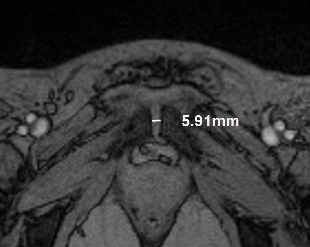
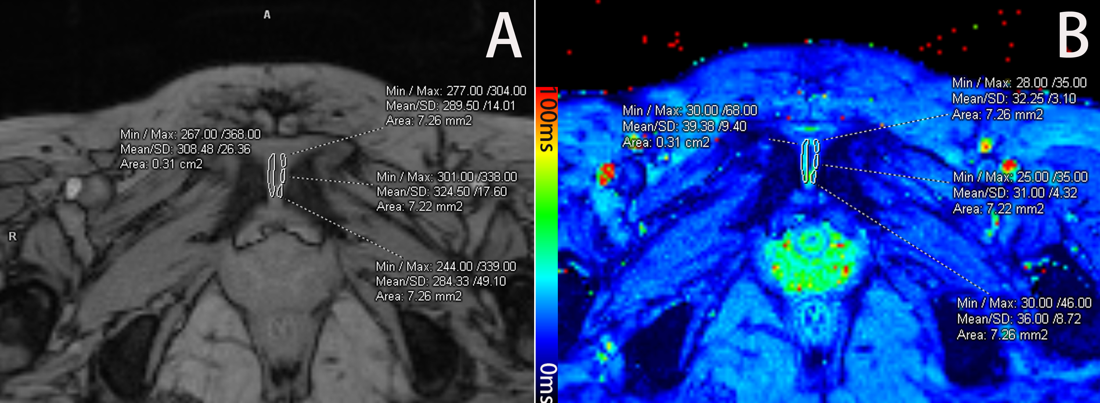
Sixty-one women during first pregnancy, 48 women during second pregnancy, and 64 nulliparous women underwent pubic symphysis MRI exam on a 1.5T MR scanner, including multi-echo T2* mapping sequence. The MRI protocols included a multi-echo gradient recalled echo sequence with five consecutive echoes for T2* mapping. The parameters were as follows: TR 423 ms, TE 4.35, 11.83, 19.31, 26.79, and 34.27 ms, flip angle 60°, bandwidth 260 Hz/pixel, field of view 320 × 320 mm2, matrix: 256 × 256, 11 slices, slice thickness 3.0 mm, voxel size 1.25 × 1.25 × 3.0 mm3, and acquisition time was 3:13 min. The T2* maps were generated using an inline processing package (Syngo MapIt; Siemens Healthcare, Erlangen, Germany), which used a log-linear, least squares method to fit the echo intensities. The slice with the narrowest pubic symphyseal width at middle part was identified, the average pubic symphyseal width and anterior pubic ligament at mid-point were then measured on TE=11.83ms images (Figure 1). The ROIs analysis was undertaken manually by one senior radiologist. Two ROIs were hand drawn on TE=11.83ms images of left and right side of cartilage, and 3 sub-regions (anterior, middle and posterior) were drawn on each side of cartilage (Figure 2) for zonal evaluation. The ROIs were positioned within the fibrocartilage zone seen in figure 2. The volume for left/right region was 28.6 ± 4.5 mm2; and the sub-region was 7.0 ± 0.8 mm2. Then the same ROIs were copied to the T2* maps to obtain T2* values. Mean values for T2* were used for statistical analysis and expressed as mean ± standard deviation (SD). T2 * values were analyzed and compared among the three groups using analysis of covariance. The average pubic symphyseal width was measured, the correlation between pubic symphyseal width and T2* values of cartilage were analyzed using Pearson's correlation coefficient.Results
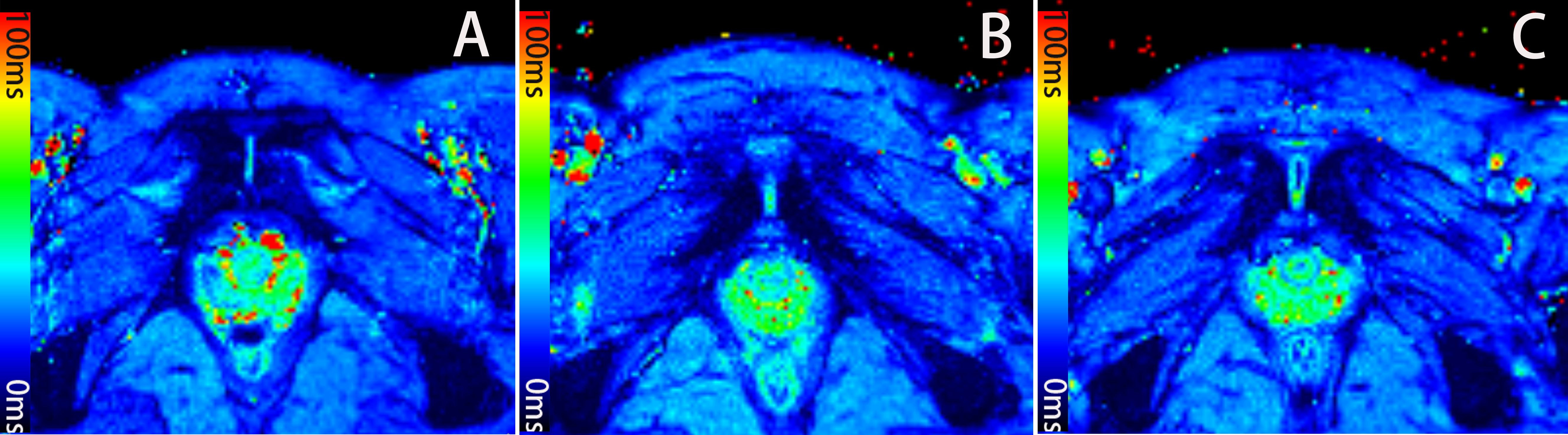
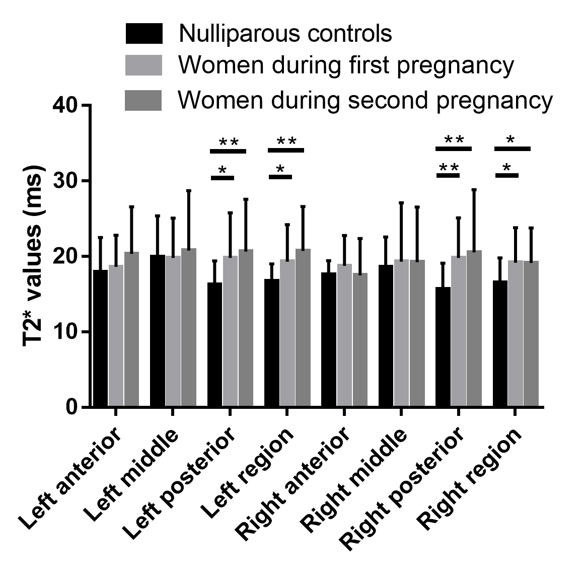
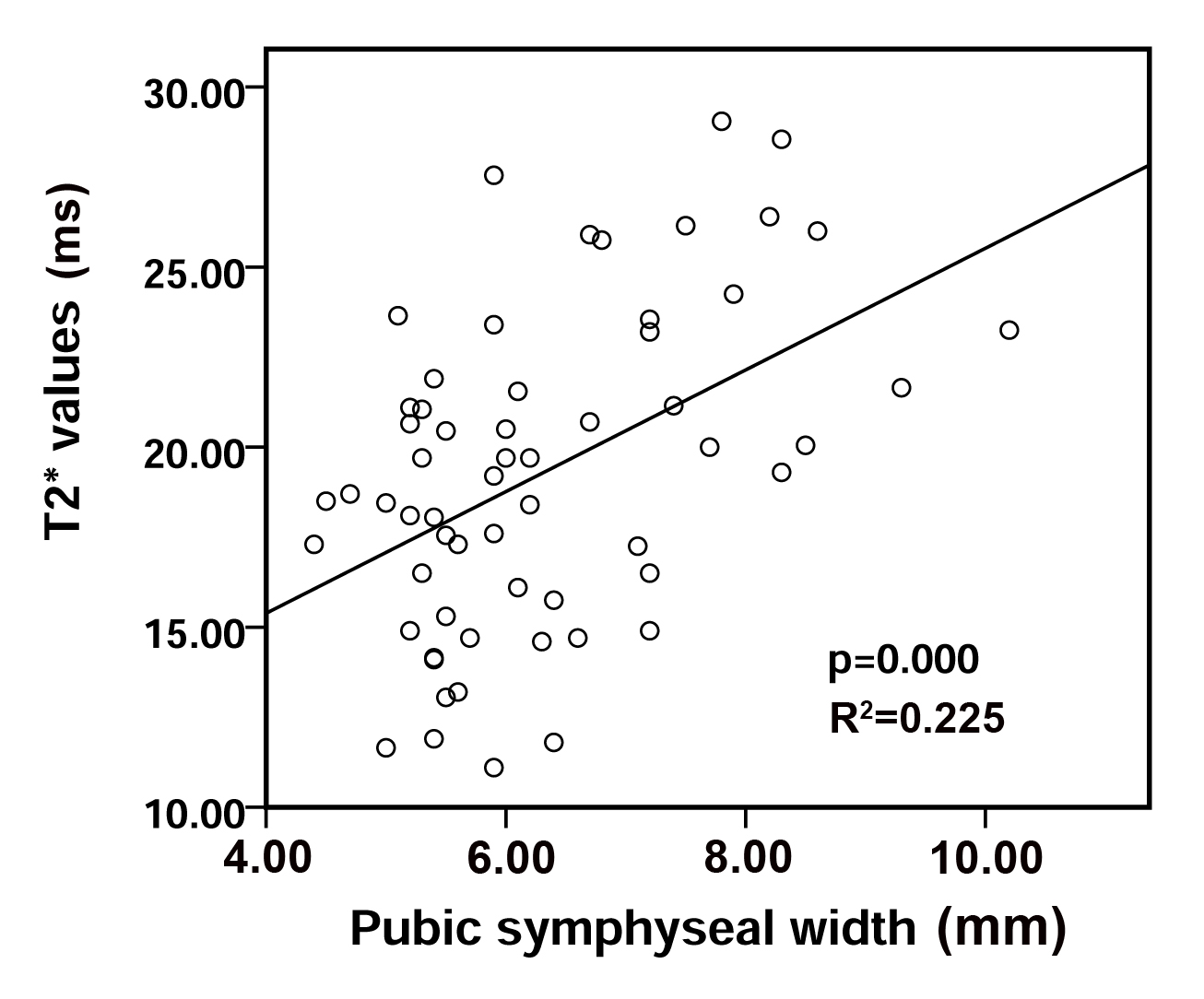
The pubic symphyseal width increased significantly during late pregnancy (p <0.001), while no differences were found between women during the first and second pregnancies (p = 0.089). Compared with nulliparous women, T2* values increased significantly in late pregnancy in left/right cartilage of pubic symphysis, the differences were mainly driven by the posterior sub-region, while no differences were found between women during first and second pregnancies (Figure 3 and 4). The positive linear correlation (Figure 5) between symphyseal width and mean T2* values of cartilage was only seen in the group of women during first pregnancy (R2=0.225, p=0.000), no correlation in the other two groups.Discussion
T2 relaxation time reflects the ability of free water proton molecules to move and to exchange energy inside the cartilaginous matrix 8. T2* mapping is feasible to display the cartilage of pubic symphysis clearly, and the statistics results showed that T2* values of cartilage increase significantly during late pregnancy. The primary reason for this alteration is the structure of pubic symphysis would change for the demand of child-birth. A hormone called relaxin released during pregnancy would increase the elasticity of tendons, ligaments, and muscles 9,10, which induce the pressure from fetus to widen the pubic gap easily and to stretch the cartilage between pubic bones. Our results verified that the pubic symphyseal width increase from ~4.8mm to ~6.2/6.7mm in first/second pregnancy. We further found a positive correlation between T2* values of cartilage and pubic symphyseal width, which was only seen in women during first pregnancy. We assumed that the alterations of cartilage structure after first pregnancy was difficult to recover completely, so the correlation was not seen in the women during second pregnancy. The sub-region matrix comparison of cartilage demonstrated that the differences in T2* values between pregnant women and nulliparous controls were mainly driven by the posterior region of each side. There are four ligaments to maintain the stability of pubic symphysis, among which posterior ligament is the thinnest one, so the posterior cartilage is the most prone to be stretched when the fetus pressed the pubic symphysis.Conclusions
T2* mapping is a sensitive quantitative method capable of detecting the pubic symphyseal cartilage changes related to pregnancy, and the T2* values increased significantly during late pregnancy.Acknowledgements
We would like to express my thanks to all the volunteers and pregnant women enrolled in this study.References
1. Becker I, Woodley SJ, Stringer MD. The adult human pubic symphysis: a systematic review. Journal of anatomy. 2010;217(5):475-487.
2. Chang EY, Ma Y, Du J. MR Parametric Mapping as a Biomarker of Early Joint Degeneration. Sports health. 2016;8(5):405-411.
3. Hontoir F, Clegg P, Nisolle JF, Tew S, Vandeweerd JM. Magnetic resonance compositional imaging of articular cartilage: What can we expect in veterinary medicine? Veterinary journal (London, England : 1997). 2015;205(1):11-20.
4. Le J, Peng Q, Sperling K. Biochemical magnetic resonance imaging of knee articular cartilage: T1rho and T2 mapping as cartilage degeneration biomarkers. Annals of the New York Academy of Sciences. 2016;1383(1):34-42.
5. Link TM, Neumann J, Li X. Prestructural cartilage assessment using MRI. Journal of magnetic resonance imaging : JMRI. 2017;45(4):949-965.
6. Niitsu M. [Articular cartilage MRI: Update]. Nihon rinsho Japanese journal of clinical medicine. 2016;74(6):924-930.
7. Krause FG, Klammer G, Benneker LM, Werlen S, Mamisch TC, Weber M. Biochemical T2* MR quantification of ankle arthrosis in pes cavovarus. Journal of orthopaedic research : official publication of the Orthopaedic Research Society. 2010;28(12):1562-1568.
8. Mamisch TC, Hughes T, Mosher TJ, et al. T2 star relaxation times for assessment of articular cartilage at 3 T: a feasibility study. Skeletal radiology. 2012;41(3):287-292.
9. Samuel CS, Butkus A, Coghlan JP, Bateman JF. The effect of relaxin on collagen metabolism in the nonpregnant rat pubic symphysis: the influence of estrogen and progesterone in regulating relaxin activity. Endocrinology. 1996;137(9):3884-3890.
10. Pinheiro MC, Moraes SG, Battlehner CN, Caldini EG, Toledo OM, Joazeiro PP. Histochemical and ultrastructural study of collagen fibers in mouse pubic symphysis during late pregnancy. Micron. 2004;35(8):685-693.
Figures