2773
The Vertebral Marrow Microstructure in Healthy Young Adults with Intravoxel Incoherent Motion Diffusion-weighted MRI1Shanxi Medical University Second Affiliated Hospital, Taiyuan, China, 2GE Healthcare, MR Research China, Beijing, Beijing, China, 3Sichuan Provincial People's Hospital, Chengdu, China, 4Shanxi Medical University, Taiyuan, China
Synopsis
Intravoxel incoherent motion diffusion-weighted MRI (IVIM) plays an important role in detecting and monitoring microstructure. It offers diffusion and perfusion information at the same time without contrast agents. In this work, we demonstrated IVIM parameters can discriminate the microstructure of marrow in younger women from men compared with the apparent diffusion coefficient (ADC) values. It may provide noninvasive methods for evaluating cellularity, vascular volume and blood velocity of bone marrow.
Purpose
To assess the microstructure of marrow using the parameters of intravoxel incoherent motion (IVIM), then to investigate gender-related cellular and capillary network in healthy young adults.Introduction
Bone marrow is the fourth largest organ of the human body. MRI has become preferred over other imaging modalities in evaluating marrow compositions. Although the cellularity of marrow was positive correlation with apparent diffusion coefficient (ADC) values, the ADC values was overlap between neoplastic marrow and normal marrow. Dynamic contrast-enhanced MRI has been applied to analyze the marrow perfusion of capillary network in patients with benign and malignant marrow diseases. It is not widely used in clinical work because of the injection of MRI tracer. IVIM provides both diffusion and perfusion quantification at the same time without contrast agents. The study of IVIM in marrow is less well established currently. Therefore, IVIM was introduced in the current study to assess the vertebral bone marrow microstructure and investigate gender-related cellular and capillary network of vertebral marrow in healthy young adults.Materials and Method
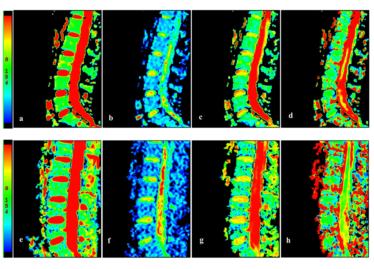
60 healthy young adults (30 men and 30 women) underwent MRI scans at 1.5 Tesla (Signa, GE Healthcare, Waukesha, WI) with routine sagittal T1-weighted sequence, conventional DWI and IVIM sequence. The applied imaging parameters are as follows: FSE sagittal T1-weighted: TR = 400.0ms, TE = 9.3ms, section thickness = 5.0mm, no gap, NEX = 4, FOV = 36cm×36cm, matrix = 320×192, acquisition time was 2 minutes and 29 seconds; conventional DWI: b=0, 500s/mm2, TR = 4000.0ms, TE = 66.5ms, section thickness = 5.0mm, no gap, NEX = 4, FOV = 36cm×28.8cm, matrix = 128×96, acquisition time was 2 minutes and 12 seconds; IVIM: b= 0, 10, 25, 50, 100, 200, 400, 600, 800, 1000, 1200s/mm2, TR = 2000ms, TE = 91ms, other parameters are the same as the DWI sequence and the acquisition time was 8 minutes and 45 seconds.Results
The D value in women was significantly higher than that in men (0.28×10⁻³mm2/s ± 0.04 vs 0.20 ×10⁻³mm2/s ± 0.04; t=-8.653, P=0.001, Table 1, Figure 1, 2). The ADC value was not statistically significant between women and men (t=-1.337, P=0.186, Table 1, Figure 1, 2). The f was significantly higher in women compared with men (31.70%±3.65 vs 28.94%±5.52; t=-2.286, P=0.026, Table 1, Figure 1, 2). However, D* was significantly lower in women 46.72×10⁻³mm2/s ± 12.90 than that 91.78 ×10⁻³mm2/s ± 30.83 in men (t=7.387, P=0.001, Table 1, Figure 1, 2). The D, f and D* values were not significant difference among the second, third and fourth lumbar vertebras of women (t=0.022, P=0.978; t=0.445, P=0.642; t=0.942, P=0.397, respectively, Table 2). There were same results in men (t=0.370, P=0.691; t=0.634, P=0.533; t=0.326, P=0.723, respectively, Table 2).Conclusion
IVIM parameters can discriminate the microstructure of marrow in younger women from men compared with the ADC values. It may provide noninvasive methods for evaluating cellularity, vascular volume and blood velocity of bone marrow.Acknowledgements
No acknowledgement found.References
[1] Vogler JB, Murphy WA, Murphy. Bone Marrow Imaging. Radiology 1988,168 (3):679-693.
[2] Moulopoulos LA, Dimopoulos MA. Magnetic resonance imaging of the bone marrow in hematologic malignancies. Blood 1997,90(6):2127-2147.
[3] Nonomura Y, Yasumoto M, Yoshimura R, Haraguchi K, Ito S, Akashi T, Ohashi I. Relationship between bone marrow cellularity and apparent diffusion coefficient. J Magn Reson Imaging 2001,13(5):757-760.
[4] Shih TT, Liu HC, Chang CJ, Wei SY, Shen LC, Yang PC. Correlation of MR lumbar spine bone marrow perfusion with bone mineral density in female subjects. Radiology 2004,233(1):121-128.
[5] Le Bihan D, Breton E, Lallemand D, Grenier P, Cabanis E, Laval-Jeantet M. MR imaging of intravoxel incoherent motions: application to diffusion and perfusion in neurologic disorders. Radiology 1986,161(2):401-407.
Figures