2770
Simultaneous evaluation of bone and cartilage status in asymptomatic FAI subjects and healthy controls using PET-MRI1Medical Imaging, The Ottawa Hospital, Ottawa, ON, Canada, 2Radiology, University of Ottawa, Ottawa, ON, Canada, 3Brain Imaging Centre, The Royal Hospital, Ottawa, ON, Canada, 4Nuclear Medicine, The Ottawa Hospital, Ottawa, ON, Canada, 5Medicine, University of Ottawa, Ottawa, ON, Canada, 6Orthopaedic Surgery, The Ottawa Hospital, Ottawa, ON, Canada
Synopsis
In this study subjects with asymptomatic FAI and healthy controls underwent PET-MRI to investigate hip bone activity (by [18F]-Sodium Fluoride (NaF) PET) and hip cartilage proteoglycan content (by MRI T1ρ mapping). The FAI subjects showed an increased [18F]-NaF uptake in the femoral head neck junction and in lateral area of the acetabulum, indicating increased bone remodelling. Hip cartilage T1ρ values were significantly higher in the anterior-lateral region for the FAI group, revealing a loss of proteoglycan with a possible connection to an early osteoarthritis stage. PET-MRI has potential to be a useful non-invasive tool to research FAI in vivo.
Introduction
Cam morphology of the femoral head-neck junction may lead to osseous conflict with the acetabular rim and may predispose to femoroacetabular Impingement (FAI)1. The impingement damages the cartilage, which can lead to the development of osteoarthristis (OA) with underlying increased bone mineral density of the acetabular rim2. In this study the uptake of [18F]-Sodium Fluoride (NaF) in the hip bones is studied simultaneously with the evaluation of the proteoglycan content of the hip cartilage (MRI T1ρ mapping) using simultaneous Positron Emission Tomography and Magnetic Resonance Imaging (PET-MRI) in asymptomatic FAI subjects and healthy controls.Methods
In this institutional review board approved study six subjects with asymptomatic cam deformity [6 males, mean age: 42.4 years (range: 34.8-51.8)] and four healthy controls [4 males, mean age: 38.9 years (range: 31.7-47.5)] were scanned in a 3T PET-MRI machine (Siemens Biograph mMR). Inclusion criteria were: No previous hip pathology/surgery and no signs of OA based on radiographic assessment. T1ρ maps were acquired bi-lateral in sagittal oblique orientation using 3D gradient echo sequence combined with a spin-lock preparation: Resolution = 0.5x0.5 mm2, slice = 3 mm, spin lock times = 0,15,30,45 ms, B1 = 500 Hz. Subjects were injected with an average 299±45 MBq of [18F]-NaF and simultaneous PET imaging was performed in one bed position in axial orientation, centered on the femoral head. Images were reconstructed using the vendor supplied 3D OP-OSEM algorithm with Dixon based MR attenuation correction. T1ρ data was processed with in-house written software, by fitting the images obtained at different TSL to a mono-exponential decay. PET Standard Uptake Values (SUVs) were calculated from a single dataset reconstructed from minutes 50 to 60 post-injection. For cartilage T1ρ analysis the joint was subdivided into an Antero-Lateral (AL) and Postero-Lateral (PL) region covering 9 mm of cartilage (3 slices) on the lateral side. SUVmax and SUVmean were both analyzed in Antero-Lateral (PET_AL) and the Postero-Lateral (PET_PL) region of the acetabulum covering 10 mm superior of the acetabulum. SUVmax were evaluated in the femoral head-neck junction of the FAI subjects and in the corresponding area for the controls. The significances of the differences were evaluated using t-tests, the correlations between T1ρ and SUVs using the Spearman correlation coefficient rho.Results
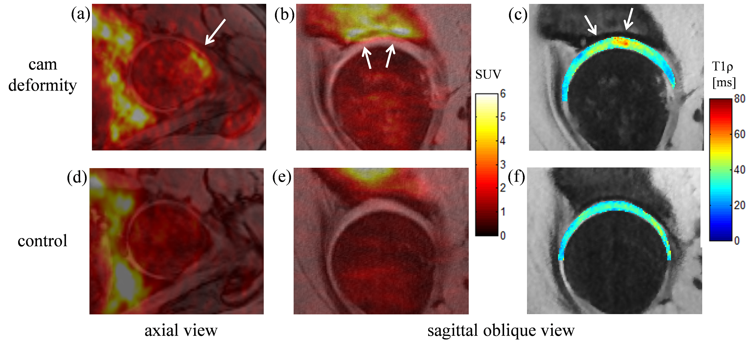
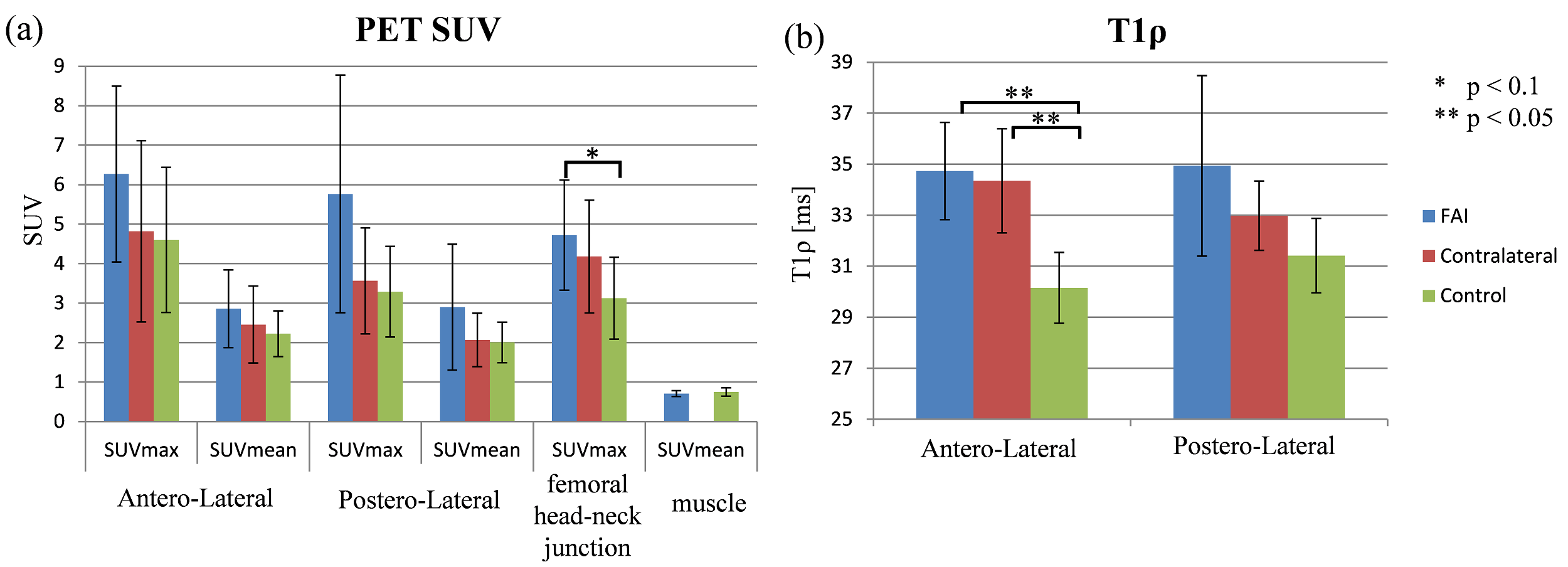
Figure 1 shows PET SUV and cartilage T1ρ maps as color-coded overlays on MR images for a FAI subject (a-c) and control (d-f). Increased [18F]-NaF uptake can be seen for the FAI subject in the femoral head neck junction and in lateral area of the acetabulum. T1ρ values are increased in the lateral region of the cartilage. In Figure 2a the PET SUVs for the different analyzed regions are shown. Increased SUVmax was measured in the AL and PL region of the acetabulum for the hips with cam deformity compared to the contra-lateral side and to the control group, although the differences were not significant (p>0.1). The hips with impingent had a significant (p<0.1) tracer uptake in femoral head-neck junction (SUVmax = 4.72±1.40) compared to the control (SUVmax = 3.13±1.04). (For comparison the SUVmean values of the muscle are shown for both groups). The cartilage T1ρ values are shown in Figure 2b. Significant increased T1ρ values were measured in the AL region for the impingement group and on the contra-lateral side compared to the control group (p<0.01). The Spearman correlation analysis between SUVmax (femoral head-neck junction) and T1ρ (AL) resulted in a moderate correlation of rho = 0.43, between SUVmax (AL) and T1ρ (AL) in no correlation (rho = 0.04).Discussion
Biochemical sensitive MRI methods, such as T1ρ mapping, have been shown to be useful tools for probing the cartilage macromolecular content in subjects with FAI3. Initial studies using [18F]-NaF PET have been successfully used to evaluate bone activity in symptomatic and asymptomatic FAI subjects4. In this study both techniques were simultaneously used to evaluate the status of the cartilage as well as the bone activity/remodelling in asymptomatic FAI subjects and controls. The increased cartilage T1ρ values in the AL region of the hip joint reveal a loss of proteoglycan with a possible connection to an early OA stage. The increased uptake of [18F]-NaF (SUVmax) in the femoral head-neck junction at the impingent side indicates an elevated bone turnover and ongoing bone remodelling. Although the increased uptake of the bone marker was not significant in the lateral area of the acetabulum, three FAI subjects had SUVmax values above 6, indicating higher bone remodelling. The moderate correlation between T1ρ (AL) and SUVmax (femoral head-neck junction) indicates the connection between bone remodelling at the impingement and loss of proteoglycans in the cartilage tissue. Although there was an increased SUVmax value in the lateral region of the acetabulum in three FAI subjects, no correlation was found between the SUVmax in the acetabulum and T1ρ AL indicating a different time phase in the FAI/OA progress.Conclusion
PET-MRI can be used to evaluate the status of cartilage (using biochemical sensitive MRI) and the bone turnover (using [18F]-NaF PET) simultaneously. It has potential to be a useful non-invasive tool to research FAI and to investigate the interaction between cartilage and bone in vivo.Acknowledgements
No acknowledgement found.References
1. Ganz R, Parvizi J, Beck M, et al. Femoroacetabular impingement: a cause for osteoarthritis of the hip. Clin Orthop Relat Res. 2003;417:112-20.
2. Beaulé PE, Grammatopoulos G, Speirs A, et al. Unravelling the hip pistol grip/cam deformity: Origins to joint degeneration. J Orthop Res. 2018;36:3125-35.
3. Anwander H, Melkus G, Rakhra KS, et al. T1ρ MRI detects cartilage damage in asymptomatic individuals with a cam deformity. J Orthop Res. 2016;34:1004-9
4. Kobayashi N, Inaba Y, Tezuka T, et al. Evaluation of local bone turnover in painful hip by 18F-fluoride positron emission tomography. Nucl Med Commun. 2016;37:399-405.
Figures