2763
Gender-related variation of IVIM diffusion weighted imaging observed in lumbar vertebral bone marrow: a healthy volunteer study
Fan Qing1, Ren Huipeng1, Wang Xiaohu1, Shen Tianbo1, Wei Xiaocheng2, and Ren Zhuanqin1
1Baoji Central Hospital, Shaanxi Province, Baoji, China, 2GE Healthcare, MR Research China, Beijing, China
1Baoji Central Hospital, Shaanxi Province, Baoji, China, 2GE Healthcare, MR Research China, Beijing, China
Synopsis
Despite the fact that diffusion MRI is well established in bone marrow as a valuable imaging modality, only a limited number of studies have investigated the intravoxel incoherent motion (IVIM) approach. In this study, bi-exponential IVIM was used to measure diffusion and perfusion fraction in lumbar vertebral bone marrow in 99 healthy subjects. The results demonstrated a gender-related variation in D and f while D* was an independent factor. Our findings may help to better understand the variation of water diffusion and perfusion imaging in lumbar vertebral bone marrow in-vivo with healthy and pathological condition.
INTRODUCTION
As a noninvasive way to probe tissue structure at a microscopic scale, Diffusion-weighted imaging (DWI) has proven to be powerful medical imaging modality1-2. Measurements of the water apparent diffusion coefficient (ADC) and tissue perfusion in vertebral bone marrow especially in lumbar provides a means for tissue microstructure characterization and it has been shown that the water diffusivity in bone marrow allows to distinguish between benign and malignant lesions3. Recently, Intravoxel Incoherent Motion (IVIM) DWI has gained extensive attention and widely used in clinical applications for various organs. IVIM could contribute to the signal attenuation observed with diffusion MR imaging, such as blood microcirculation in the capillary networks (perfusion) and not only molecular diffusion4, without need of contrast agents administration. However, to date, only a very limited number of IVIM studies have been performed on bone marrow and little attention has been paid to the feature on IVIM parameters including diffusion coefficient(D), pseudodiffusion coefficient (D*) and perfusion fraction (f)5-6. Here we systematically quantify diffusion and perfusion in the lumbar vertebral bone marrow with IVIM in healthy volunteers to investigate the character of diffusion and perfusion parameters.METHODS
99 volunteers with 64 males (age range 18–87 years, mean age 57.45 ± 15.4years) and 35 females (age range 18–74 years, mean age 55.8 ± 10.4 years) joined this study with written informed consents after the approval of the local Ethics Committee. All data were acquired on a 3-T whole-body human MRI scanner (Discovery 750W, GE Healthcare, Milwaukee, WI, USA) with an 8-channel phase array spine coil. The IVIM multi b images were acquired in sagittal plane with a reduced field of view diffusion sequence. Detailed parameters are: TR/TE = 4000/73.2 ms; FOV = 260 mm × 104 mm; matrix size = 128 × 52; NEX = 2; slice thickness = 4 mm; scan time = 4 min and 56 s. b values = 0, 50, 100, 150, 200,400, 600, 800 and 1000 s/mm2, respectively. After acquisition, images are transferred to vendor provided advanced workstation for post processing. IVIM parametric maps including D, D* and f were calculated using bi‑exponential fitting by the segmented fitting method. Statistical analysis was conducted by independent-samples T test on L1-L5 vertebral between different genders.RESULTS
Table 1 shows the value of D, D* and f from L1 to L5 in different genders. Over all speaking, the average value of D for all lumbar vertebral was significantly higher (p<0.01) in male than female; while the average value of f was lower (p<0.01). On the other hand, D* did not demonstrate gender dependency(p=0.16). Specifically, the value of D from L1-L3 and were significantly higher (p<0.01, p<0.01 and p<0.05 respectively) in male subjects, while the value of f from L1-L2 were significantly lower (p<0.05). There were no significant differences of D from L4-L5, f from L3-L5 and D* across all lumbar vertebral. The box plots of D, D* and f in the vertebral bone marrow were shown in Fig. 1.DISCUSSION and CONCLUSION
In the current study, the IVIM approach allows for measuring diffusion and perfusion fraction in vertebral bone marrow and establish a baseline with 99 healthy volunteers. We implemented a spine IVIM protocol to investigate the character of parameters D, D* and f. The most important finding of this study was that we found a gender-related variation in D and f while D* was an independent factor, which might originate from the variation of water-fat composition of vertebral bone marrow and balance with a relatively steady blood microcirculation. Despite the fact that quantitative MRI is well established in bone marrow and has great potential as a tool which allowing earlier detection, diagnosis, staging, and monitoring of disease progression or response to therapy, only a limited number of studies have used the IVIM approach6-8. These studies revealed a relatively broad range of perfusion fraction value 9-10 which is consistent with our findings. The gender-related variation of IVIM parameters observed in this study may help us find a better way to understand the variation of the water diffusion and perfusion imaging in lumbar vertebral bone marrow in vivo with healthy and pathological condition.Acknowledgements
References
- Koutoulidis V, Papanikolaou N, Moulopoulos LA. Functional and molecular MRI of the bone marrow in multiple myeloma. Br J Radiol. 2018;91: 20170389.
- Sunghoon Park, Kyu-Sung Kwack, Nam-Su Chung et al. Intravoxel incoherent motion diffusion-weighted magnetic resonance imaging of focal vertebral bone marrow lesions: initial experience of the differentiation of nodular hyperplastic hematopoietic bone marrow from malignant lesions. Skeletal Radiology. May 2017, Volume 46, Issue 5, 675–683.
- Biffar A, Dietrich O, Sourbron S, Duerr HR, Reiser MF, Baur-Melnyk A. Diffusion and perfusion imaging of bone marrow. Eur J Radiol. 2010 Dec;76(3):323-8.
- A.J.Marchand, E.Hitti, F.Monge et al. MRI quantification of diffusion and perfusion in bone marrow by intravoxel incoherent motion (IVIM) and non-negative least square (NNLS) analysis. Magnetic Resonance Imaging. Volume 32, Issue 9, November 2014, 1091-1096.
- Chen Ye, Daoyun Xu, Yongbin Qin, Estimation of intravoxel incoherent motion parameters using low b-values. PLoS One. 2019; 14(2): e0211911.
- Hui Tan, Hui Xu, Feifei Luo et al. Combined intravoxel incoherent motion diffusion-weighted MR imaging and magnetic resonance spectroscopy in differentiation between osteoporotic and metastatic vertebral compression fractures. Journal of Orthopaedic Surgery and Research. (2019) 14:299.
- Denis Le Bihan. What can we see with IVIM MRI? NeuroImage. 187 (2019) 56–67.
- Mami Iima, MD, Denis Le Bihan. Clinical intravoxel incoherent motion and diffusion MR imaging: Past, Present, and Future. Radiology.Volume 278: Number 1-January 2016.
- Ma W, Wei M, Han Z et al. The added value of intravoxel incoherent motion diffusion weighted imaging parameters in differentiating high-grade pancreatic neuroendocrine neoplasms from pancreatic ductal adenocarcinoma. Oncol Lett. 2019 Nov;18(5):5448-5458.
- Li YT, Cercueil JP, Yuan J et al. Liver intravoxel incoherent motion (IVIM) magnetic resonance imaging: a comprehensive review of published data on normal values and applications for fibrosis and tumor evaluation. Quant Imaging Med Surg. 2017 Feb;7(1):59-78.
Figures
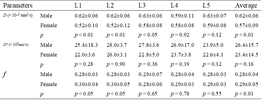
Mean and SD of D, D*and f for different gender from L1-L5.
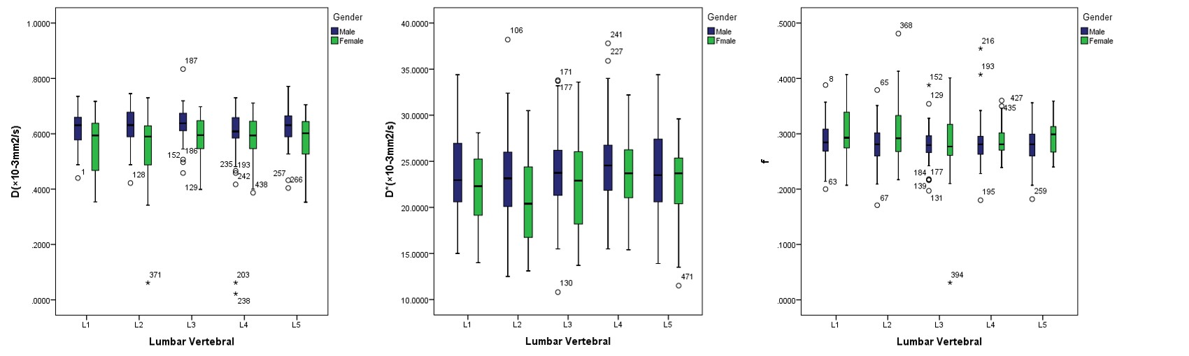
The
value of D, D*
and f
in vertebral bone marrow, obtained from the
IVIM measurements and averaged over all volunteers.