2760
Detection of femoral head ischemia using DCE-MRI in Children with Slipped Capital Femoral Epiphysis (SCFE)1Radiology, Chiba Children's Hospital, Chiba, Japan, 2Center for Frontier Medical Engineering, Chiba University, Chiba, Japan, 3Orthopedics, Chiba Children's Hospital, Chiba, Japan
Synopsis
In this study, we reveal a scheme to evaluate the femoral head ischemia using optimized DCE-MRI for preoperative diagnosis in children with unstable SCFE. The consistency of the evaluation of DCE-MRI is verified through compare the cases which were classified as ischemia or normal after surgery. As a result, optimized DCE-MRI is a useful tool to detected femoral head ischemia in children with unstable SCFE, which allows doctors to plan a treatment strategy ahead of time. PEI and MTE using DCE-MRI contribute to an objective evaluation of the perfusion status.
Purpose
Slipped capital femoral epiphysis (SCFE) is one of the hip joint lesions in children during the period of growth. A treatment of unstable SCFE in particular is difficult, since many patients could have developed a complication such as avascular necrosis (AN) becoming the factor of a poor function outcome and early osteoarthritis (OA).1 It is difficult to predict an occurrence of postoperative AN since there is no specific methods to evaluate the patient's status before the surgery. However, it is important for patients to reveal the preoperative perfusion status such as ischemia or congestion in femoral head in order to prevention a complication.2,3 Recently, we reported the optimization of dynamic contrast-enhanced magnetic resonance imaging (DCE-MRI) to evaluate the femoral head perfusion status in children.4 This method apply to evaluate perfusion status in unstable SCFE.In this study, we reveal a scheme to evaluate the femoral head ischemia using optimized DCE-MRI for preoperative diagnosis in children with unstable SCFE. The consistency of the evaluation of DCE-MRI is verified through compare the cases which were classified as ischemia or normal after surgery.Materials and Methods
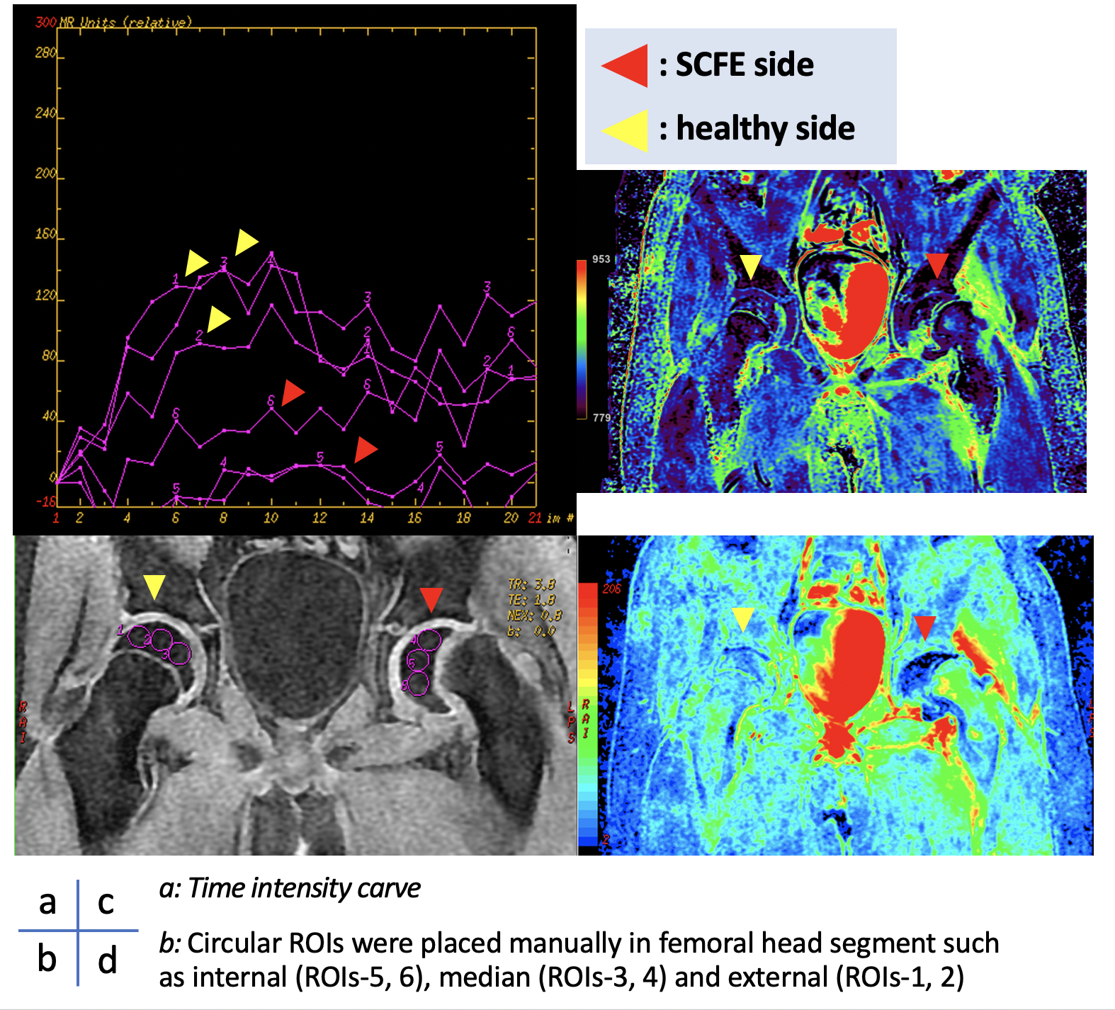
Eleven pediatric patients with unstable SCFE before surgery were examined in this study (9 males, 2 females; mean age, 11.3 years). All subjects were scanned using a 1.5 Tesla MRI scanner. The perfusion imaging with optimized DCE-MRI sequence was done from 10 minutes immediately after the administration of a gadolinium contrast agent. The perfusion status was evaluated by calculating a color map using positive enhanced integral (PEI) and mean time to enhance (MTE), and by calculating the time intensity curve (TIC) of both the healthy and SCFE-affected femoral heads. (Figure A) Before the surgery, PEI and MTE ratio were calculated between the healthy side and SCFE-affected side in femoral heads (Figure B). All subjects were classified into two groups such as normal or ischemia after the surgery. The ischemia group was compared to the normal group for preoperative PEI and MTE ratio.Results & Discussions
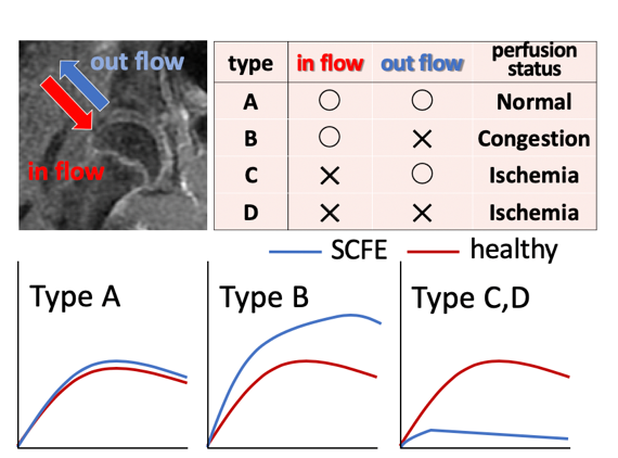
Nine out of all patients were classified as a normal group and the other two patients were classified as an ischemia group. PEI and MTE ratio were significantly lower in the ischemia group than the normal group.(PEI ratio: 0.32 ± 0.08 versus 2.01 ± 0.79, MTE ratio: 0.96 ± 0.79*10-2 versus 1.02 ± 2.16*10-2). P-values were obtained from the Wilcoxon ranksum test (*P<0.05). (Figure D) There were four types of predicted perfusion status from DCE-MRI (Figure C). Therefore, nine out of all patients were classified as the perfusion status of type B, as the slipped epiphysis caused impaired venous perfusion. Moreover, the other patients were classified as the perfusion status of type C or D, as the slipped epiphysis caused ischemia. Figure E shows the clinical images which detected a blood flow disorder affected by SCFE. The perfusion color mapping using DCE-MRI allows observers to do an objective evaluation since it calculates color maps, such as PEI and MTE, as well as TIC types.Conclusion
Optimized DCE-MRI is a useful tool to detected femoral head ischemia in children with unstable SCFE, which allows doctors to plan a treatment strategy ahead of time. PEI and MTE using DCE-MRI contribute to an objectiveevaluation of the perfusion status.Acknowledgements
No acknowledgement found.References
1. Novais EN. et al. Clin Orthop Relat Res. 2012. Dec;470(12):3432-8.
2. Staatz G. et al. Eur Radiol. 2007. Jan; 17:163-168
3. Rhoad R.C. et al. J Pediatr Orthop. 1999. Mar-Apr;19:164-168
4. Kojiro O, et al. ISMRM2018 #5160.
Figures
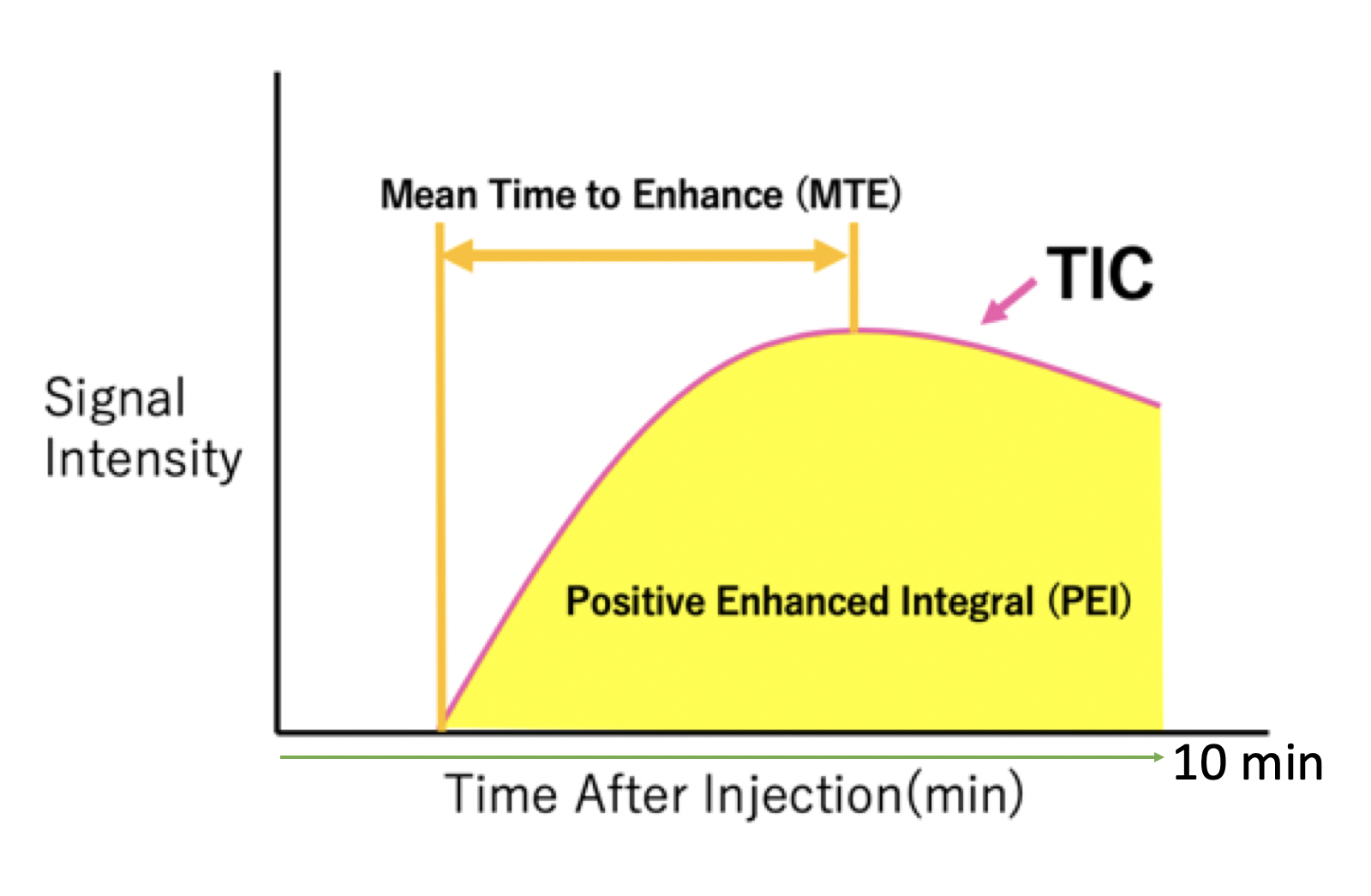
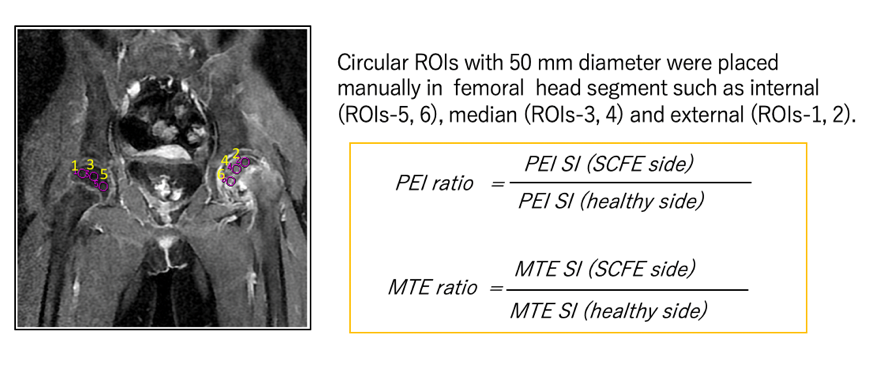
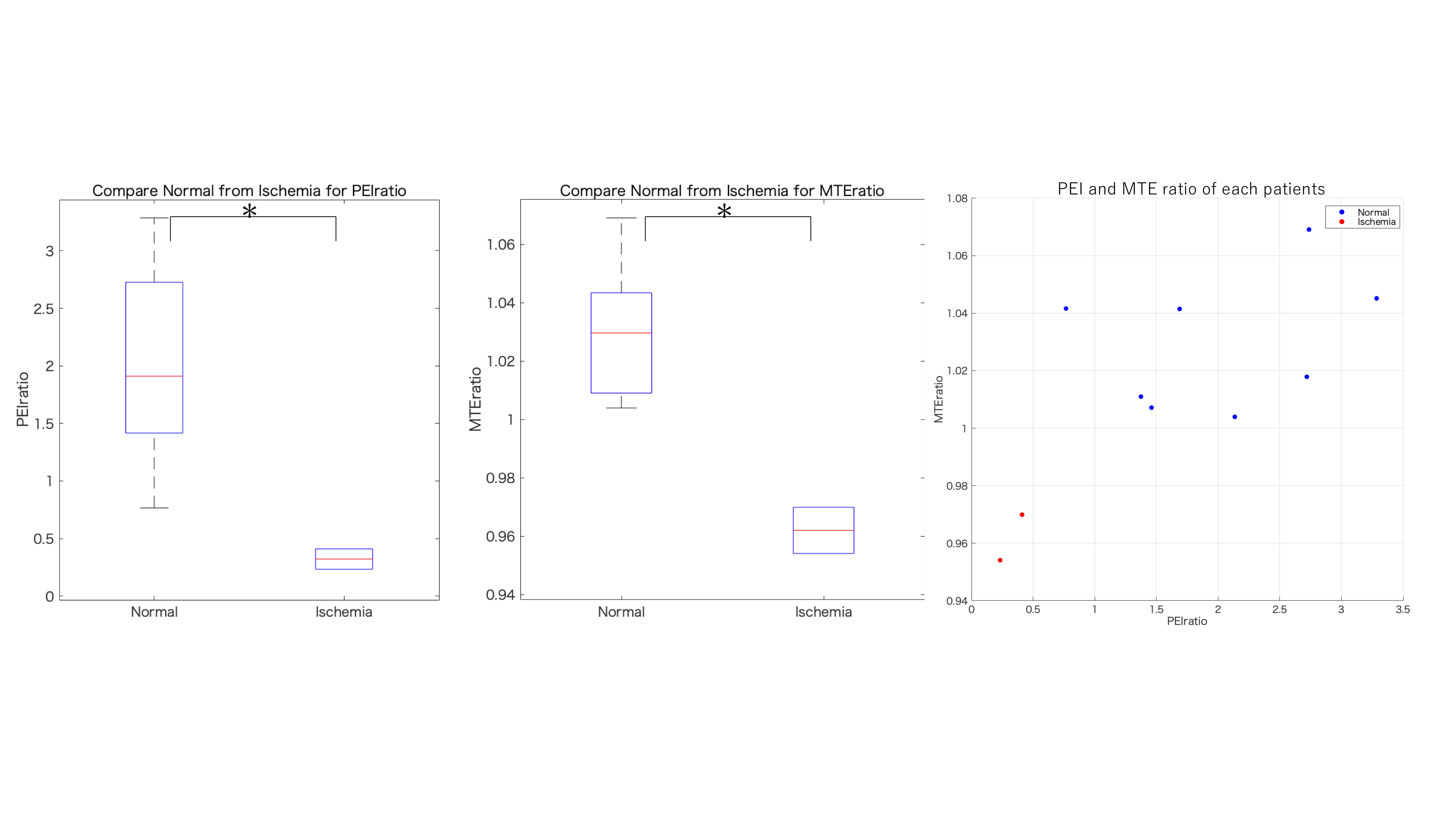
Figure C: Comparison with signal ratio between ischemia group and normal group