2748
T2 Relaxation Time Reveals Improvement of Articular Cartilage Quality After Bariatric Surgery at 12-month Follow-up1Research Unit of Medical Imaging, Physics and Technology, University of Oulu, Oulu, Finland, 2Medical Research Center, University of Oulu and Oulu University Hospital, Oulu, Finland, 3Department of Diagnostic Radiology, Oulu University Hospital, Oulu, Finland, 4Center for Life Course Health Research, University of Oulu, Oulu, Finland, 5Finnish Institute of Occupational Health, Oulu, Finland, 6Department of Surgery, Oulu University Hospital, Oulu, Finland, 7Department of Physical Medicine and Rehabilitation, Oulu University Hospital, Oulu, Finland
Synopsis
Obesity has become a worldwide phenomenon with nearly tripling since 1975: 13% of adults over 18 are obese. We set out to study articular cartilage of knee joint in obese individuals using T2 relaxation time. Our study population underwent Roux-en-Y gastric bypass operation and were compared with a control group of obese individuals in a 12-month follow-up. Our results suggest improved quality of cartilage in the lateral compartment of femoral cartilage after bariatric surgery.
Introduction
Multiple studies have demonstrated that osteoarthritis of knee is accelerated by obesity1. It is commonly hypothesized that at least a part of the cartilage injury is a result of altered mechanical stress due to obesity2.We set out to study obese subjects’ knee cartilage using T2 relaxation time, which is known to correlate with cartilage structure and mechanical properties3. The study population underwent Roux-en-Y gastric bypass operation4, and had a 12-month T2 measurement follow-up of knee articular cartilage. The findings were compared with a conservative care control group of obese subjects. Our aim was to assess the effect of gastric bypass on the knee articular cartilage, evaluated using T2 relaxation time measurement.
Methods
The study group consisted of 48 subjects (40 females) aged from 32 to 68 (mean 49.4±7.8) years with BMI ranging from 32.2 to 54.9 (mean 40.7±6.5) kg/m2. The participants were on waiting list for bariatric surgery at Oulu University Hospital. The study group underwent Roux-en-Y gastric bypass operation. The control group consisted of 33 subjects (27 females) aged from 29 to 65 (mean 50.4±8.9) years with BMI ranging between 34.0 and 53.7 (mean 40.5±4.9) kg/m2. This non-surgical group consisted of subjects who were treated with conservative weight reduction regimens in local health care centers.All subjects underwent imaging of knee joint with a 3.0 T MRI unit (Skyra, Siemens Healthcare, Erlangen, Germany) using a 18-channel body matrix coil and a 32-channel spine coil. Sagittal 2D T2 mapping was acquired using a MESE sequence (TR=1680 ms; TEs=13.8, 27.6, 41.4, 55.2, 69 ms; FOV=160x160 mm2; matrix=256x256; resolution=0.62x0.62x3.00 mm3). Each study participant was scanned prior to surgery and at 12 month post-operatively.
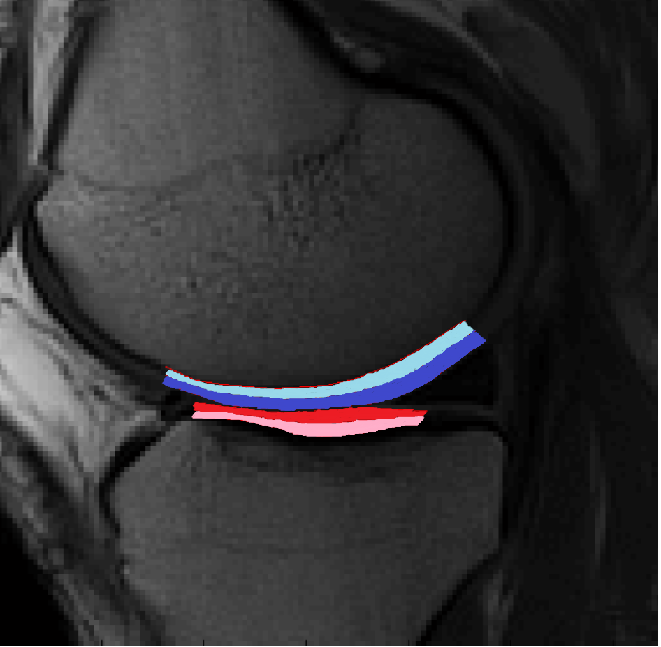
T2 relaxation time maps were calculated pixel-wise using monoexponential model fitting and mean T2 values were obtained by segmenting sagittal T2 maps of knee joint. Cartilage of central femur (cF) and central tibia (cT) of medial (M) and lateral (L) compartments were chosen to represent weight bearing regions of the knee joint (Figure 1).
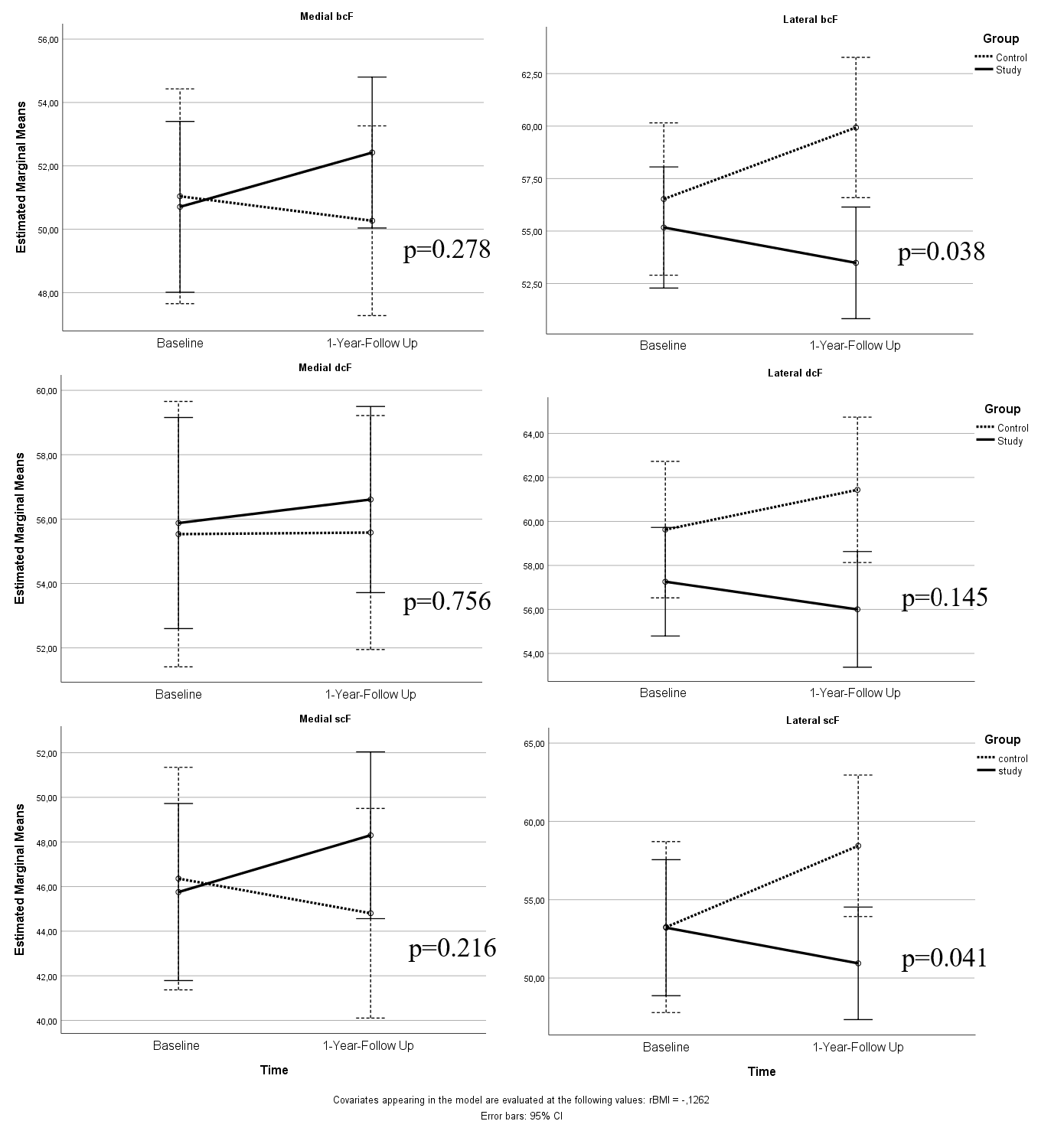
Statistical analysis was performed using SPSS 26.0. Repeated measures analysis of variance was conducted to evaluate the influence of gastric bypass surgery on the T2 relaxation times of different regions of knee cartilage. The analyses were adjusted for relative BMI change (rBMI) and KL-grade. Analysis was performed for each region on bulk (b, full-thickness), deep (d, 50% lower half) and superficial (s, 50% upper half) partitions of the cartilage.
Results
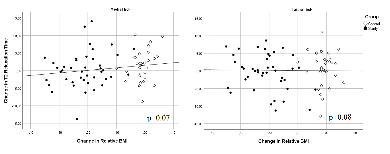
Mean change of BMI in the intervention group was -9.1±3.3 kg/m2 and in the control group -0.3±1.3 kg/m2 (p< 0.01 between the groups). The gastric bypass operation decreased the T2 relaxation time in the lateral compartment of femoral cartilage (p-values for bcF, dcF and scF were 0.038, 0.145 and 0.041, respectively). No significant differences were observed in the medial side of femur or either side of the tibia. Results for femur are presented in Figure 2. Dependency of the change in rBMI and change in T2 value for medial (p=0.07) and lateral (p=0.08) bcF are presented in Figure 3.Discussion
We observed a decrease in T2 relaxation time values in the study group and increasing values in the lateral compartment of femoral cartilage of the control group. Previously, elevation of T2 has been associated with degeneration of the collagen network and increase in tissue hydration, and thus, decreased cartilage quality3,5. Therefore, our results suggest that gastric bypass surgery may have a positive effect on the lateral compartment of femoral cartilage already after 12 months. No differences were observed in the medial femoral or tibial compartments. Another 12-month follow-up study suggested that weight loss is associated with increased cartilage dGEMRIC index, reflective of increased proteoglycan content, and decreased knee cartilage loss in the medial compartment but not in the lateral compartment6. In our 12-month follow-up we observed a similar trend with decreased T2 relaxation times due to weight loss in the medial compartment, although not statistically significant. However, we did not see any significant differences in T2 in medial compartment in our study group. This could be due to the different quantitative methods used to assess cartilage: dGEMRIC reflects proteoglycan content, whereas T2 reflects collagen content4, and these constituents may be differently affected in degeneration. We also speculate that after losing weight and being able to move around more frequently, the mechanical loading pattern of the knee joint could be altered, which may result in altered mechanical stress of the knee. A recent study reported a lower T2 increase rate in both knee compartments after 96 months of weight loss with diet and exercise, suggesting a slower progression of cartilage degeneration with weight loss programs7. In our study, obese subjects who underwent bariatric surgery experienced improvements in knee cartilage quality, as reflected by significantly decreased T2 in lateral compartment, already after 12 months. It remains to be seen, whether T2 decrease of the medial compartment will eventually catch up with the lateral compartment over a longer follow-up period.Conclusion
Our results suggest that gastric bypass may improve the quality of lateral femoral cartilage. The results also demonstrate similarities with previous findings suggesting a trend towards improvement of medial femoral cartilage quality with weight loss.Acknowledgements
No acknowledgement found.References
1. Pottie P, Presle N, Terlain B, Netter P, Mainard D, Berenbaum F. Obesity and osteoarthritis: more complex than predicted! Ann Rheum Dis 2006;65(11):1403-5
2. Vincent HK, Heywood K, Connelly J, Hurley RW. Obesity and weight loss in the treatment and prevention of osteoarthritis. PM R. 2012 May;4(5 Suppl):S59-67. doi: 10.1016/j.pmrj.2012.01.005.
3. Nissi MJ, Töyräs J, Laasanen MS, et al. Proteoglycan and collagen sensitive MRI evaluation of normal and degenerated articular cartilage. J Orthop Res. 2004 May;22(3):557-64.
4. Buchwald H, Avidor Y, Braunwald E, Jensen MD, Pories W, Fahrbach K, Schoelles K. Bariatric surgery: a systematic review and meta-analysis. JAMA. 2004 Oct 13;292(14):1724-37.
5. Baum T, Joseph GB, Arulanandan A, et al. Association of magnetic resonance imaging-based knee cartilage T2 measurements and focal knee lesions with knee pain: data from the Osteoarthritis Initiative. Arthritis Care Res (Hoboken). 2012 Feb;64(2):248-55. doi: 10.1002/acr.20672.
6. Anandacoomarasamy A, Leibman S, Smith G, et al. Weight loss in obese people has structure modifying effects on medial but not on lateral knee articular cartilage. Ann Rheum Dis. 2012 Jan;71(1):26-32. doi: 10.1136/ard.2010.144725.
7. Gersing AS, Schwaiger BJ, Nevitt MC, et al. Weight loss regimen in obese and overweight individuals is associated with reduced cartilage degeneration: 96-month data from the Osteoarthritis Initiative. Osteoarthritis Cartilage. 2019 Jun;27(6):863-870. doi: 10.1016/j.joca.2019.01.018.
Figures