2740
Site-Specific Quantitative µMRI and Polarized Light Microscopy (PLM) Study of Young Rabbit Femur Cartilage
Yang Xia1, Syeda Batool1, and Mohammad Hammimi1
1Physics, Oakland University, Rochester, MI, United States
1Physics, Oakland University, Rochester, MI, United States
Synopsis
This work aims to characterize the site-specific and depth dependent molecular and morphological structures in young rabbit femur cartilage, using quantitative µMRI (T2 relaxation) and Polarized light microscopy (PLM).
Introduction
Articular cartilage (AC) is a thin layer of protective tissue covering the articular ends of bones in diarthrodial joints. It is a load-bearing tissue with complex composition and architecture. The molecular composition of AC mainly are water, proteoglycans (PGs), and collagen network. The architecture of AC is dominated by its collagen network, which has a depth-dependent organization. In mature cartilage, an arcade-like fibril structure exists, with a 90˚ orientation difference between the surface fibers and the deep fibers. It has been found that arcade-like structure is absent in juvenile or inborn cartilage.Material and methods
A number of 12-14 weeks old White New Zealand male rabbits were used in this study. 3 different sample sites were chosen on femoral medial condyle (anterior, central and posterior). One cartilage-bone plug (~2.0 mm thick) was harvested from each location, with the full thickness of cartilage still attached to underlying bone. Quantitative T2 mapping of the specimens was carried out at various orientations in B0 using a magnetization-prepared 2D spin-echo sequence at slice thickness 0.8 mm and at 9.75 µm/pixel resolution. After µMRI, 6.0 µm thin sections were cut from each sample to generate quantitative PLM images at 1 µm resolution. In addition, thin histological sections that spanned the entire central femoral medial condyles (Fig 1a) were imaged at 4.0 µm/pixel resolution, to study the heterogeneity of the collagen fibril organization across the tissue in both horizontal and vertical directions.Results
We found significant topographical variations in both cartilage thickness and its collagen organization across the joint surface at different anatomical sites.µMRI Results: AC in T2-weighted images of medial central and medial posterior samples appeared bilaminar at the 0˚ and its corresponding T2 profiles had an asymmetrical bell-shaped curve that resembles the classical three-zone structure in the tissue (Fig 1a). AC in T2-weighted image of anterior site appeared less laminated at 0° and its T2 profile was broader and symmetrical along the depth. The T2 anisotropy effect was smaller in the anterior site as compared to central and posterior sites.
PLM results: The medial central and medial posterior sites have developed the arcade like structure but the anterior sites are not - the collagen fibers there run predominantly parallel to articular surface and there is increased cellular density at the anterior site (Fig 1b). The quantitative retardation and azimuthal profiles for all three sites has been obtained and compared. Overall, the lowest retardation values (which indicate the lowest mutual organization among the collagen fibers) have been found in the medial anterior site, with the predominant parallel fibers w-r-t to articular surface. The PLM results from the entire central femoral medial condyles (Fig 2) clearly support the µMRI and PLM results from the individual blocks.
Discussion
T2 relaxation time is sensitive to PG and water molecules interactions and exhibit strong anisotropy, based on organizational anisotropy of the collagen matrix in tissue. The depth-dependent T2 profiles at various orientations w-r-t main magnetic field for all three sites reflect the variation of molecular dynamics modulated by the fibril organization in tissue. The present report demonstrates the site specific changes of collagen fiber organization and molecular dynamics during growth and maturation. The probable explanation for these differences along the same joint surface could be either variable growth rate, pattern of bone epiphysis or exposure to different mechanical loading conditions at different anatomical sites of same joint. In terms of function and structure relationships, the collagen network arrangement is responsible for lateral expansion of cartilage tissue during compression. Hence the collagen fiber orientation is critically important to understand this mechanism. It has been suggested that during growth and maturation period musculoskeletal system can be intervened at early age in order to improve the collagen mechanical properties, and our findings of site specific differences in the collagen network development among different sites on a single joint surface will be useful to design further techniques and developments in fields like cartilage tissue engineering in specific joints.Conclusion
Since the collagen network organization plays a significant role in managing the mechanical properties of articular cartilage, our findings in the site specific differences in collagen network organization can be useful to improve the understanding of site specific mechanical properties and mechanobiology of tissue in growth and maturation.Acknowledgements
NIH R01 grant (AR 69047)References
1-Xia, Yang, Jonathan B. Moody, Hisham Alhadlaq, and Jiani Hu. “Imaging the Physical and Morphological Properties of a Multi-Zone Young Articular Cartilage at Microscopic Resolution.” Journal of Magnetic Resonance Imaging 17, no. 3 (2003). 2-Julkunen, P. et al. Maturation of collagen fibril network structure in tibial and femoral cartilage of rabbits. Osteoarthritis and Cartilage 18, 406–415 (2010).Figures
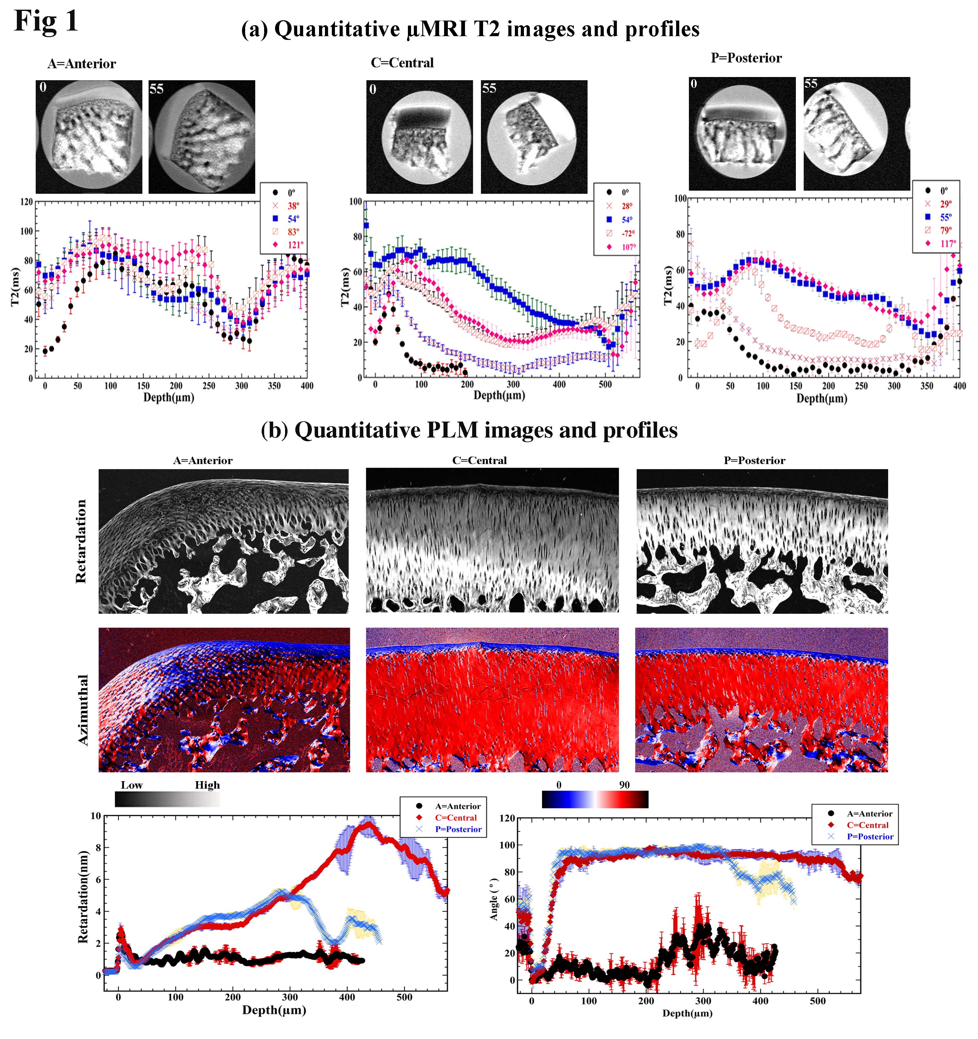
Fig 1 µMRI and PLM results of three topographical sites on the femoral condyle
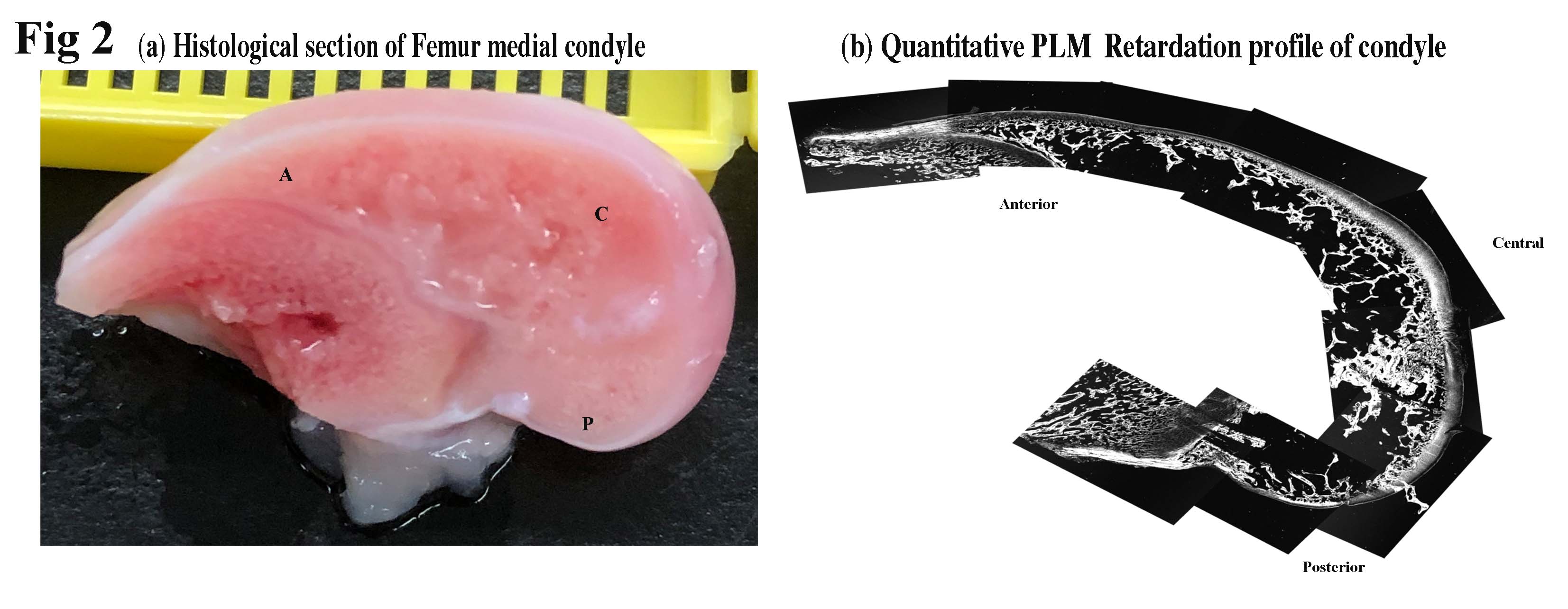
Fig 2 Histology and PLM results of the
entire central femoral medial condyle