2734
MP2RAGE improves robustness of 3D T1-Mapping of Hip Cartilage compared to Conventional Dual-Flip-Angle Acquisition1Diagnostic, Interventional and Pediatric Radiology, University Hospital Bern, Bern, Switzerland, 2Dept of Radiology, Boston Children`s Hospital, Boston, MA, United States, 3Dept of Orthopaedic Surgery, Boston Children`s Hospital, Boston, MA, United States, 4Advanced Clinical Imaging Technology, Siemens Healthcare, Lausanne, Switzerland, 5Dept of Radiology, Lausanne University Hospital and University of Lausanne, Lausanne, Switzerland, 6LTS5, École Polytechnique Fédérale de Lausanne (EPFL), Lausanne, Switzerland, 7Siemens Healthcare, Zuerich, Switzerland
Synopsis
Although commonly used for T1 mapping of hip cartilage, 3D dual flip angle (DFA) techniques are sensitive to flip angle variations related to B1 inhomogeneities. Therefore, the IR-based MP2RAGE technique was compared with a standard 3D DFA acquisition including B1 field mapping for T1 mapping of hip cartilage in a phantom study and in healthy volunteers at 3T. For the phantom study, a 2D IR-based sequence was additionally acquired to provide reference T1 maps. MP2RAGE resulted in more accurate T1 values with less regional variations compared to DFA method and therefore enables a more robust T1 mapping of hip cartilage.
Introduction
Due to the advent of femoroacetabular impingement and the almost exponential rise in joint-preserving procedures performed over the last 15 years, there has been an increasing desire to improve surgical decision in patients eligible for hip preservation surgery1. Reliable prognostic tools to identify those patients who benefit most from surgical intervention are needed2. While standard morphologic imaging has proven to be efficient in diagnosing labrum tears3, the evaluation of early cartilage damage has been elusive4. Delayed gadolinium-enhanced MRI of cartilage (dGEMRIC) as a biomarker for cartilage quality has been frequently used in clinical studies5,6 to study cartilage quality in early degenerative hip disease. Currently, the standard technique for post-contrast T1 mapping of hip cartilage is based on a 3D dual-flip-angle (DFA) gradient echo based method7. However, this technique is susceptible to B1-related flip angle variations increasing at higher B08. More recently, the magnetization-prepared 2 rapid gradient echo (MP2RAGE) sequence was introduced to obtain 3D T1 maps with minimized effect of B1 inhomogeneity even at higher B09. In this study we compared the 3D MP2RAGE technique and the standard 3D DFA acquisition against a 2D IR technique in phantoms with varying T1 values. Additionally, we assessed the variability in pre-contrast T1 of cartilage and periarticular muscles of MP2RAGE in comparison to the DFA method in healthy volunteers.Methods
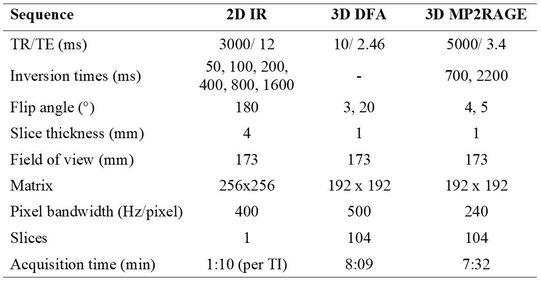
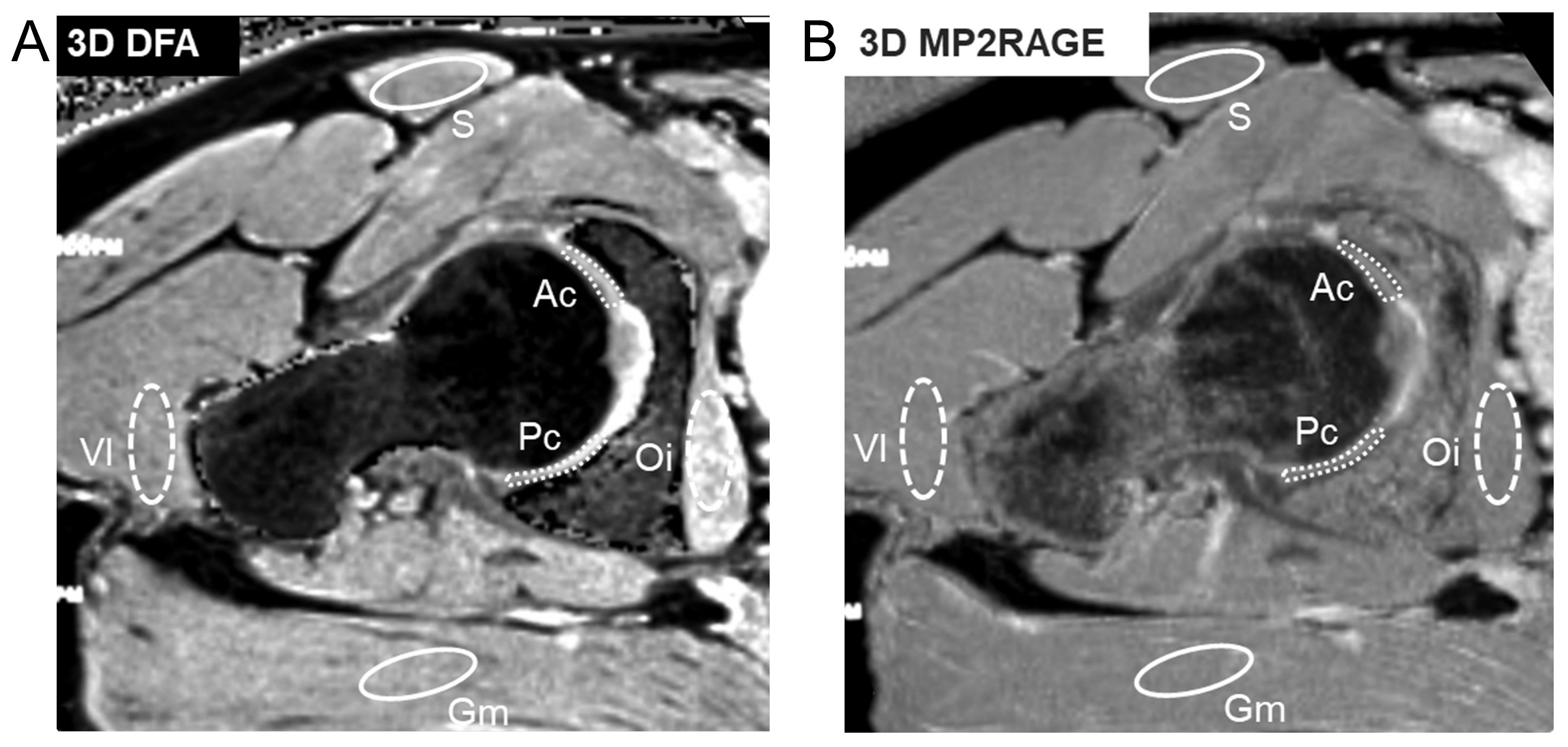
All measurements were performed at 3T (MAGNETOM Skyra, Siemens Healthcare, Germany) with a 15-channel knee coil. 3D DFA, a prototype 3D MP2RAGE, and 2D single-slice fast spin echo IR (as a reference for T1 values) sequences were acquired in a phantom study. Eleven phantoms with different concentrations of Gd-DTPA (Magnevist, Berlex Laboratories, New Jersey, USA) ranging from 0.14 to 1 mM have been used to obtain a range of T1 values. A B1-mapping sequence was acquired to assess a potential correlation between B1 variations and variations of the DFA and the MP2RAGE T1 values from the reference 2D IR T1 values. ROIs were placed for each phantom to measure T1 for the DFA, MP2RAGE, and 2D IR methods as well as B1 variations. 18 asymptomatic hips in 9 volunteers (27±2 years) underwent non-contrast, quantitative T1 imaging with the acquisition of axial-oblique MP2RAGE and DFA (including a prescan for B1 correction) images. To optimize the accuracy of T1 measurements within a range of expected cartilage pre- and post-contrast T1-values between 400 and 1100 ms, the inversion times and flip angle values for both MP2RAGE images were determined according to the simulations as described by Marques9. To reduce acquisition time, the in-plane phase-encoding direction was switched to the inner loop of the gradient echo block, and the slice-encoding including partial Fourier was set to the outer loop. Sequence parameters are summarized in Fig.1. For radial reformatting of 3D images the greater trochanter serves as landmark for the 12 o’clock position. ROIs at clock-face positions 1 o’clock–5 o’clock, 12 o’clock–7 o’clock were clustered as mean anterior and posterior cartilage regions. For measurement of periarticular muscle T1 manually placed regions of interest of comparable size in the satorius- and gluteus maximus muscles (antero-posterior) and the internal obturator- and the vastus lateralis muscle (medio-lateral) (see Fig.2).Results
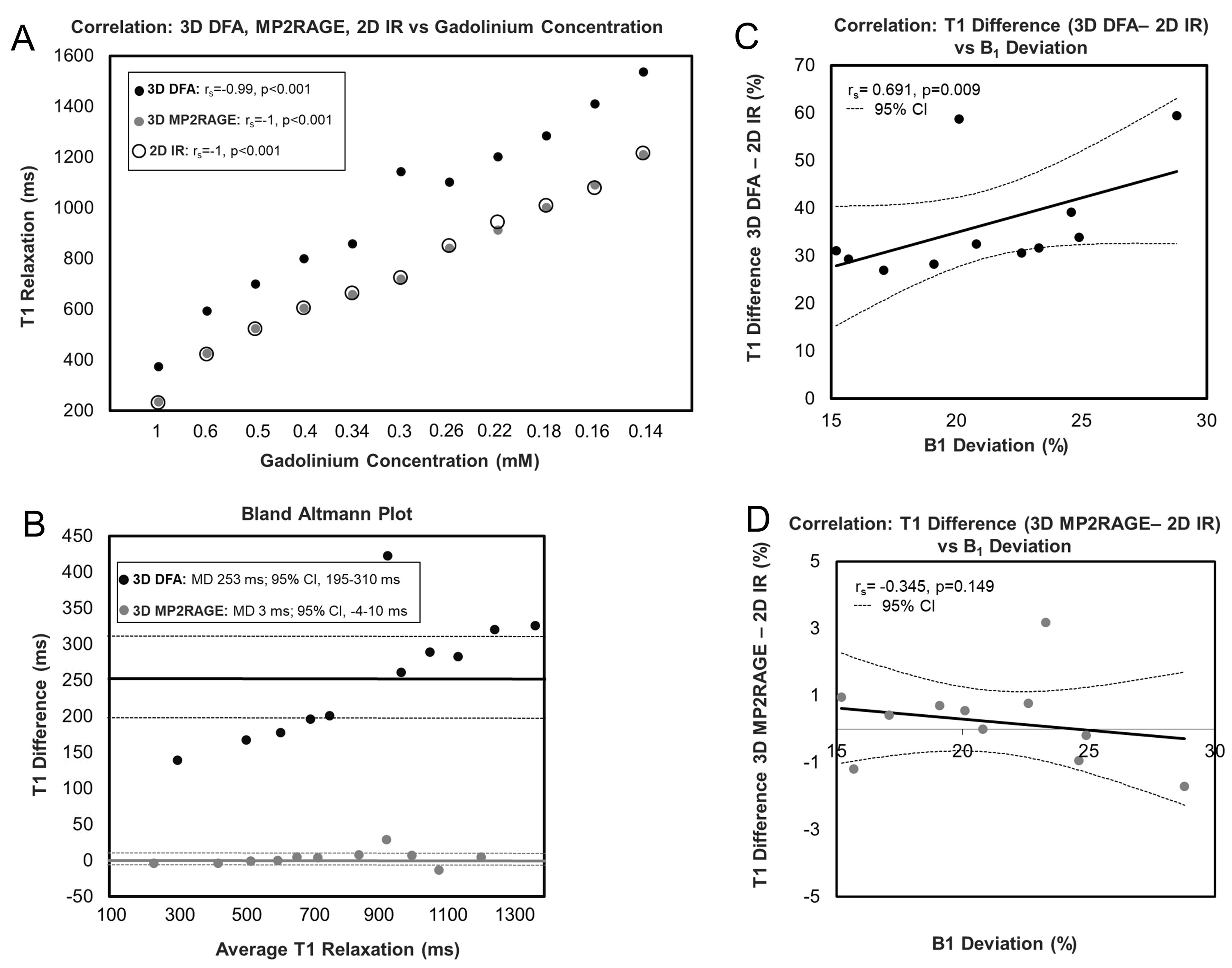
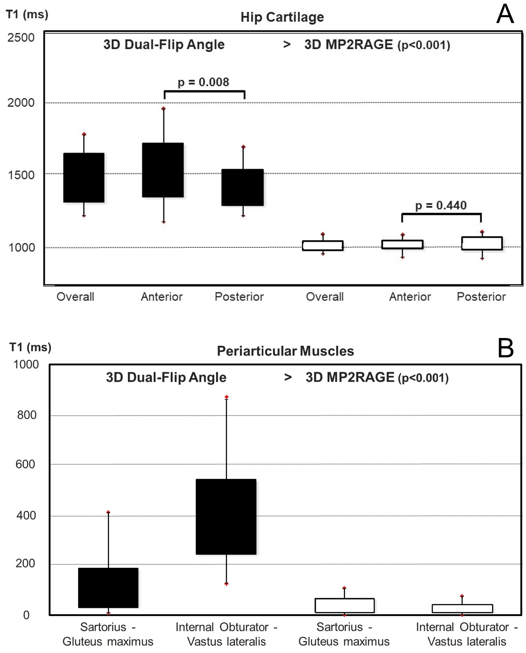
The phantom study is summarized in Fig.3. T1 values over Gd concentration in 11 phantoms reveals excellent agreement for MP2RAGE and 2D IR techniques, whereas DFA shows clear discrepancies compared to 2D IR (3A). Mean differences for the DFA- and the MP2RAGE technique compared against the 2D IR technique were 253±85 and 3±11 ms (3B). The Bland-Altman analysis showed a strong tendency of increasing T1 deviation of the DFA technique relative to the 2D IR technique with increasing T1 relaxation of the phantoms. Mean B1 variation was 21.1±4.2% (15.2%–28.8%). The discrepancy in T1 relaxation between the DFA- and the 2D IR technique increased strongly with increasing B1 variation (3C). By contrast, no such tendency was observed for MP2RAGE (3D). The volunteer study is summarized in Fig.4. Mean T1 values (range of means, 1434–1542 ms) of overall-, anterior and posterior cartilage were consistently higher (all p<0.001) for the DFA technique compared to the MP2RAGE technique with mean values ranging from 1028 to 1036 ms, respectively (4A). Regional difference between mean anterior and posterior cartilage T1 was observed for the DFA technique (106±163 ms, 95%-CI 32–185 ms, p=0.008). No such difference was observed for the MP2RAGE technique (4B). Mean T1 values (1279 to 1686 ms) of periarticular muscles were consistently higher (all p<0.001) for the DFA technique compared to MP2RAGE (1137 to 1179 ms).Discussion
Despite the used B1 prescan, inter-individual differences in T1 values of cartilage were greater with the DFA method compared to the MP2RAGE due to the greater flip-angle variations at 3T. The results supports the initial hypothesis that an IR-based acquisition of 3D T1 maps of hip cartilage using the MP2RAGE would be more robust than the B1-sensitive variable-flip-angle technique at 3T. Future studies should use intravenous or intra-articular contrast injection and compare post-contrast T1 relaxation of hip cartilage against a 2D IR T1 reference in patients with hip pain and different stages of radiographic joint degeneration.Acknowledgements
No acknowledgement found.References
1. Sutter R, Pfirrmann CWA: Update on Femoroacetabular Impingement: What Is New, and How Should We Assess It? Semin Musculoskelet Radiol 2017; 21:518–528.
2. Hanke MS, Steppacher SD, Anwander H, et al. What MRI Findings Predict Failure 10 Years After Surgery for Femoroacetabular Impingement? Clin Orthop Relat Res 2017; 475:1192–1207
3. Sutter R, Zubler V, Hoffmann A, et al.: Hip MRI: how useful is intraarticular contrast material for evaluating surgically proven lesions of the labrum and articular cartilage? AJR Am J Roentgenol 2014; 202:160–169.
4. Blankenbaker DG, Ullrick SR, Kijowski R, et al.: MR arthrography of the hip: comparison of IDEAL-SPGR volume sequence to standard MR sequences in the detection and grading of cartilage lesions. Radiology 2011; 261:863–871.
5. Schmaranzer F, Haefeli P, Hanke M, et al.: How does the dGEMRIC index change after surgical treatment for FAI? A prospective controlled study: Preliminary results. Clin Orthop Relat Res 2017; 475:1080–1099.
6. Palmer A, Fernquest S, Rombach I, et al.: Diagnostic and prognostic value of delayed Gadolinium Enhanced Magnetic Resonance Imaging of Cartilage (dGEMRIC) in early osteoarthritis of the hip. Osteoarthr Cartil 2017; 25:1468–1477.
7. Sur S, Mamisch TC, Hughes T, Kim Y-J: High resolution fast T1 mapping technique for dGEMRIC. J Magn Reson Imaging 2009; 30:896–900.
8. Siversson C, Chan J, Tiderius C-J, et al.: Effects of B1 inhomogeneity correction for three-dimensional variable flip angle T1 measurements in hip dGEMRIC at 3 T and 1.5 T. Magn Reson Med 2012; 67:1776–1781.
9. Marques JP, Kober T, Krueger G, et al. MP2RAGE, a self bias-field corrected sequence for improved segmentation and T1-mapping at high field. Neuroimage 2010; 49:1271–1281.
Figures