2712
Quantification of Fat Fraction in Muscles Adjacent to Lumbosacral Plexus within Healthy People Using IDEAL-IQ Sequence1Radiology, The First Affiliated Hospital of Nanjing Medical University, Nanjing, China, 2GE Healthcare, MR Research China, Beijing, China
Synopsis
Many diseases can result in increased fatty infiltrations within muscles adjacent to lumbosacral plexus. However, the normal level of fat content in paraneural muscles is not clear within healthy people. In this study, we applied iterative decomposition of water and fat with echo asymmetry and least-squares estimation intelligent quantification (IDEAL-IQ) to measure fat fraction (FF) of paraneural muscles within healthy participants. We found that FF values of paraneural muscles in healthy people depend on gender and age. Therefore, both factors of gender and age need to be taken into account, when assessing the FF levels of muscles in diseases.
Introduction
Lumbosacral plexus is a complex neural network composed of the ventral divisions of the coccygeal and sacral and lumbar nerves. Paraneural muscle includes gluteus maximus, gluteus medius, gluteus minimus and multifidus. Many diseases such as lumbar disc herniation, lumbosacral fracture and neoplastic diseases can result in increased fatty infiltrations within paraneural muscles1. There have been several studies confirming that the composition of muscles on the side of the disc herniation is different from the opposite with more fatty infiltrations2. However, the normal level of fat content in muscles close to lumbosacral plexus is not clear within healthy people.IDEAL-IQ, as a relatively novel MRI technique for accurate fat quantification, has been reported to assess pancreatic fat content and to evaluate fat fraction of both parotid glands and submandibular glands3, 4.
Therefore, the aim of the present study was to investigate the normal fat fraction (FF) of muscles adjacent to lumbosacral plexus in healthy people. To achieve this goal, IDEAL-IQ imaging technique was applied.
Materials and Methods
SubjectsOur prospective study was approved by the clinical research ethics board. There were 178 participants (102 men, 48.49±16.31 years; age range, 13-81 years; 76 women, 51.03±16.86 years; age range, 18–84 years) enrolled in our study and conducted an MR examination with IDEAL-IQ imaging technique.
The exclusion criteria included: (1) history of lumbar disc herniation or lumbosacral plexus abnormality; (2) spinal deformity; (3) history of vertebral or disc surgery; (4) history of tumors; (5) contraindications for MR examination.
MRI experiment
All experiments were performed at GE 3.0 T 750W with phase-array chest-body coils. Routine MRI and IDEAL-IQ technique were applied. The scan parameters for IDEAL-IQ measurement were showed at Table 1. The parametric fat fraction mapping was then automatically obtained.
Data analysis
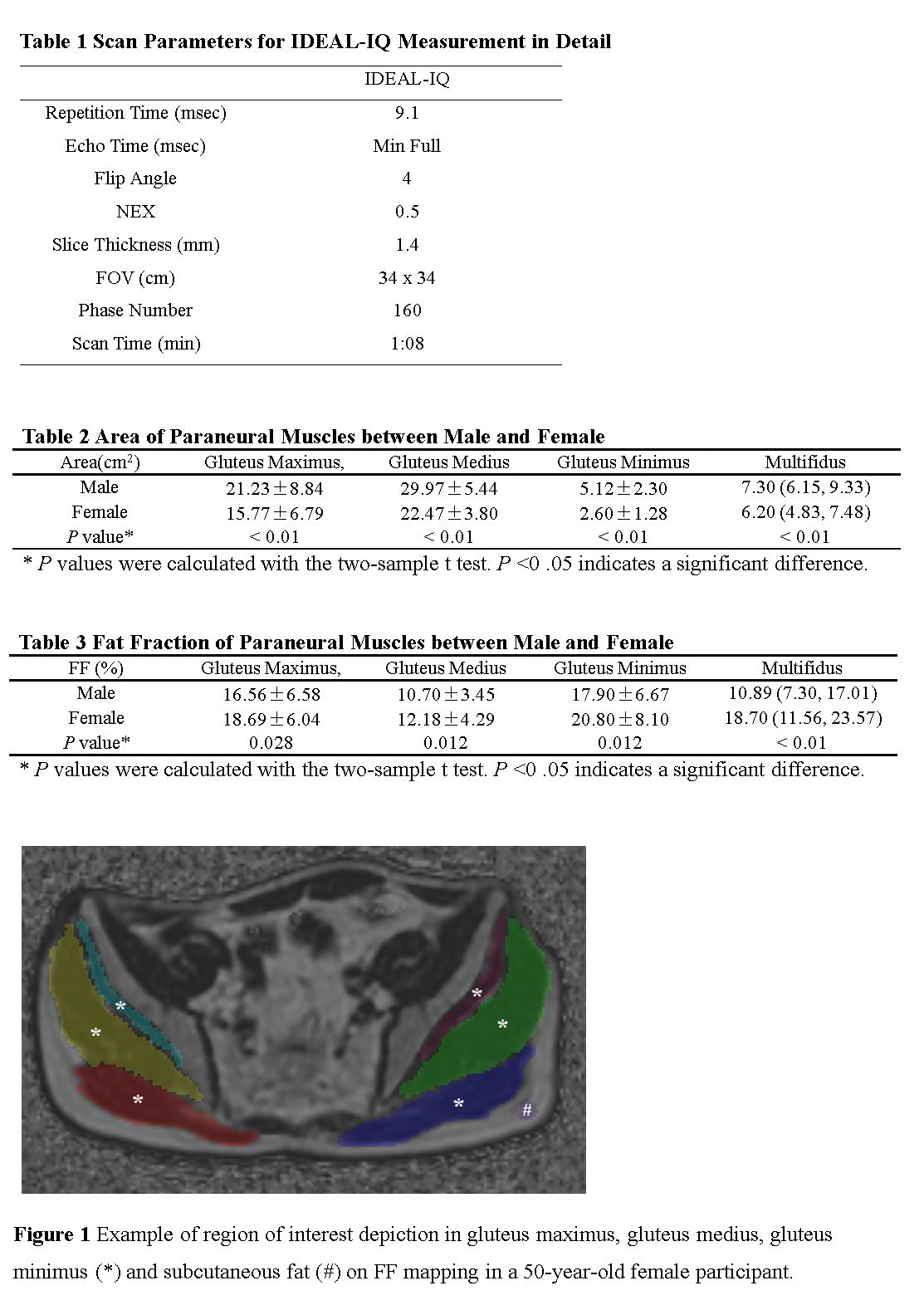
All data were analyzed at a GE MR workstation (Advantage workstation 4.6; GE Medical Systems). Region of interest (ROI) was manually depicted on FF maps from IDEAL-IQ (Figure 1). The delineation of gluteus maximus, gluteus medius and gluteus minimus were at the level of S1 nerve emerged from the sacral foramen, while the multifidus was at the level of L5 nerve emerged from intervertebral foramen. Another ROI was depicted on subcutaneous fat (area: 0.5-0.6cm2). The ratio of muscle fat fraction to subcutaneous fat fraction was recorded as FFr.
All statistical analyses were performed in SPSS software. Paired t test was performed to compare the difference of size and fat fraction between bilateral muscles. Wilcoxon rank sum test and t test were used to analyze the area and FFr of muscles. The correlations between muscles and age or gender were analyzed by Spearman’s correlation analyses. Significance threshold was set as P<0.05.
Results
No side-to-side differences were found in area and FFr of bilateral muscles (P>0.05). The area and FFr of gluteus maximus, medius and minimus followed a normal distribution, while multifidus didn’t. The corresponding mean (± SD) area of gluteus maximus, gluteus medius and gluteus minimus were 18.90±8.46, 26.77±6.07 and 4.05±2.30 (cm2). The mean (± SD) FFr of these muscles were 17.47±6.42, 11.33±3.89 and 19.13±7.43 (%).Significant differences on area and FFr of each muscle were observed between male and female healthy participants (P<0.05) (Table 2 and 3). All muscle areas of male were larger than those of female. In contrast, the FFr values of male were smaller than those of female.
We divided all the participants into three different groups according to age (13-39 years, n = 55; 40-59 years, n = 68; 60-84 years, n = 55). The area and FFr values of muscles were significantly different (P < 0.01) across three age groups in both genders. Significant positive correlations were found between age and FFr in gluteus maximus, gluteus medius, gluteus minimus and multifidus (gluteus maximus: r = 0.195, P < 0.05; gluteus medius: r = 0.324, P < 0.05; gluteus minimus: r = 0.256, P < 0.05; multifidus: r = 0.353, P < 0.05). There was no significant correlation between age and area of muscles (P > 0.05).
Discussion
The IDEAL-IQ imaging technique provides an accurate assessment of FFr. For healthy participants, bilateral muscles were usually under the same stress, so they were symmetrical in size and composition of muscles.In our study, we found that male muscles had a larger size and a lower FFr value than female. It might result from a higher muscle ratio and a higher basal metabolism in male participants than that in female. On the other hand, estrogen in female may also play a role. Meanwhile, the FFr showed a tendency to increase along with age. The decrease of exercise in elder and age-related degeneration might be the reason for it. However, exercise habit was not included in our study. Further study could take exercise as one of the factors.
Conclusion
In conclusion, our study demonstrated that the FFr values of muscles adjacent to lumbosacral plexus in healthy people depend on gender and age. Therefore, both factors of gender and age need to be taken into account, when IDEAL-IQ imaging is applied to assess the FFr levels of muscles in diseases.Acknowledgements
No acknowledgement found.References
1. Schafer AL, Vittinghoff E, Lang TF, et al. Fat infiltration of muscle, diabetes, and clinical fracture risk in older adults. J Clin Endocrinol Metab. 95(11). United States,2010. E368-72.
2. Fortin M, Lazáry À, Varga PP, et al. Paraspinal muscle asymmetry and fat infiltration in patients with symptomatic disc herniation. Eur Spine J. 25(5). Germany,2016. 1452-1459.
3. Chen Y, Long L, Jiang Z, et al. Quantification of pancreatic proton density fat fraction in diabetic pigs using MR imaging and IDEAL-IQ sequence. BMC Med Imaging. 19(1). England,2019. 38.
4. Su GY, Wang CB, Hu H, et al. Effect of laterality, gender, age and body mass index on the fat fraction of salivary glands in healthy volunteers: assessed using iterative decomposition of water and fat with echo asymmetry and least-squares estimation method. Dentomaxillofac Radiol. 48(3). England,2019. 20180263.
Figures