2711
Is fractal dimension value of the lumbar multifidus muscle associated with low back pain?1The Affiliated Hospital of Shaanxi University of Chinese Medicine, Xian Yang, China, 2MR Scientific Marketing Specialist Diagnostic Imaging Healthcare Greater China, Xi'an, China
Synopsis
Magnetic resonance imaging (MRI) is considered the best imaging method to evaluate the multifidus degeneration. The existing evaluation methods based on fat infiltration fraction (FSF) cannot fully reflect the muscle integrity and functional status. In order to obtain a tool for objective and continuous grading of multifidus degeneration. Fractal method was used to analyze the signal heterogeneity of multifidus. The fractal dimension of lumbar multifidus muscle(LMM) is closely related to LBP. The fractal dimension may be a reliable imaging marker for monitoring the degree of multifidus degeneration in symptomatic patients.
Introduction
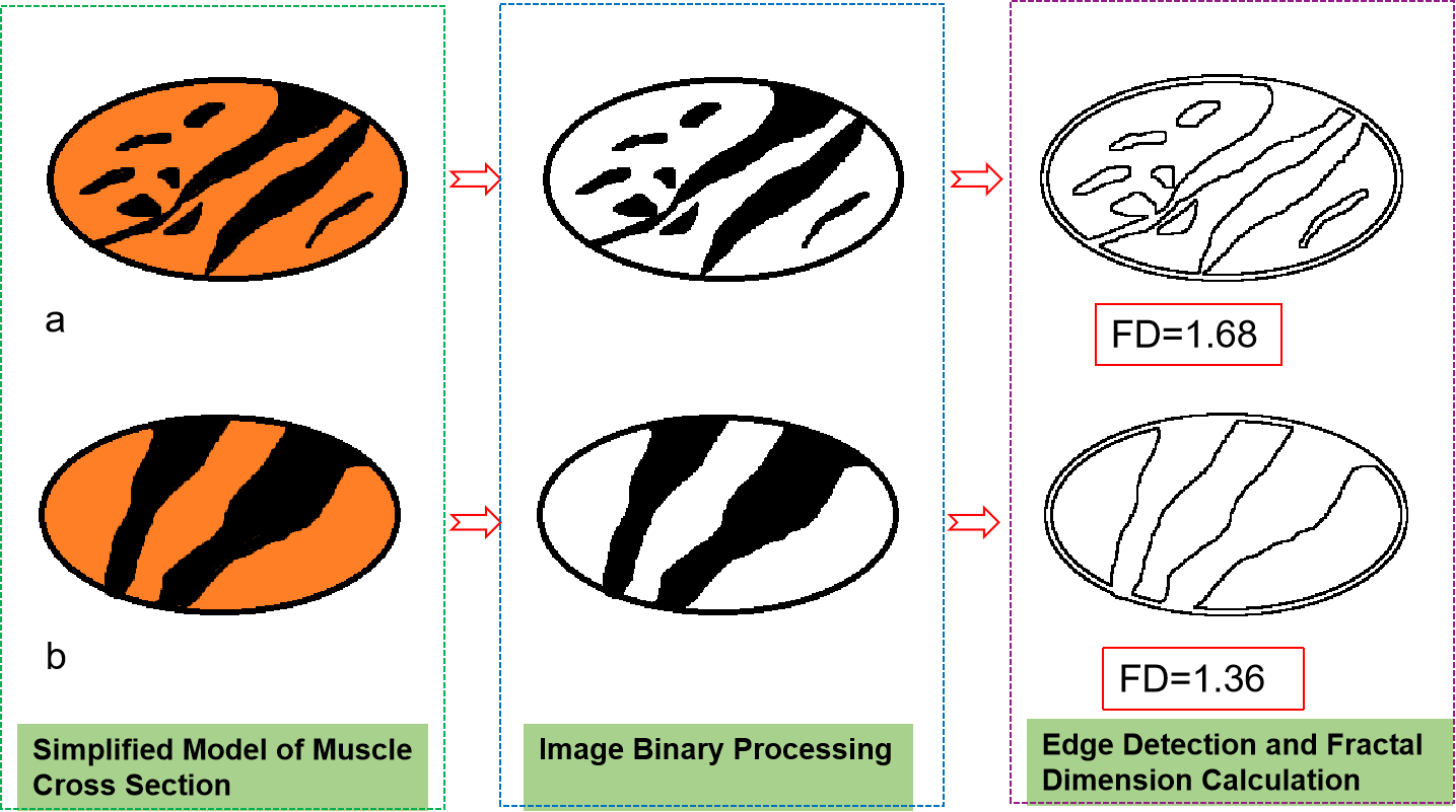
Muscle dysfunction is generally believed to be associated with low back pain (lbp). Fat infiltration fraction (FSF) have been used by many researchers as imaging biomarkers to assess muscle function and integrity1, but this is not enough to explain all (As shown in Figure 1). It is of great clinical significance to explore a quantitative method for decoding muscle integrity. Fractal dimension is an important texture features and has a strong correlation with the observer's subjective evaluation of image roughness or irregularity2. The purpose of this study is to explore the feasibility of fractal dimension decoding of multifidus degeneration, and further explore whether the fractal dimension value of multifidus is significantly related to low back pain.Methods
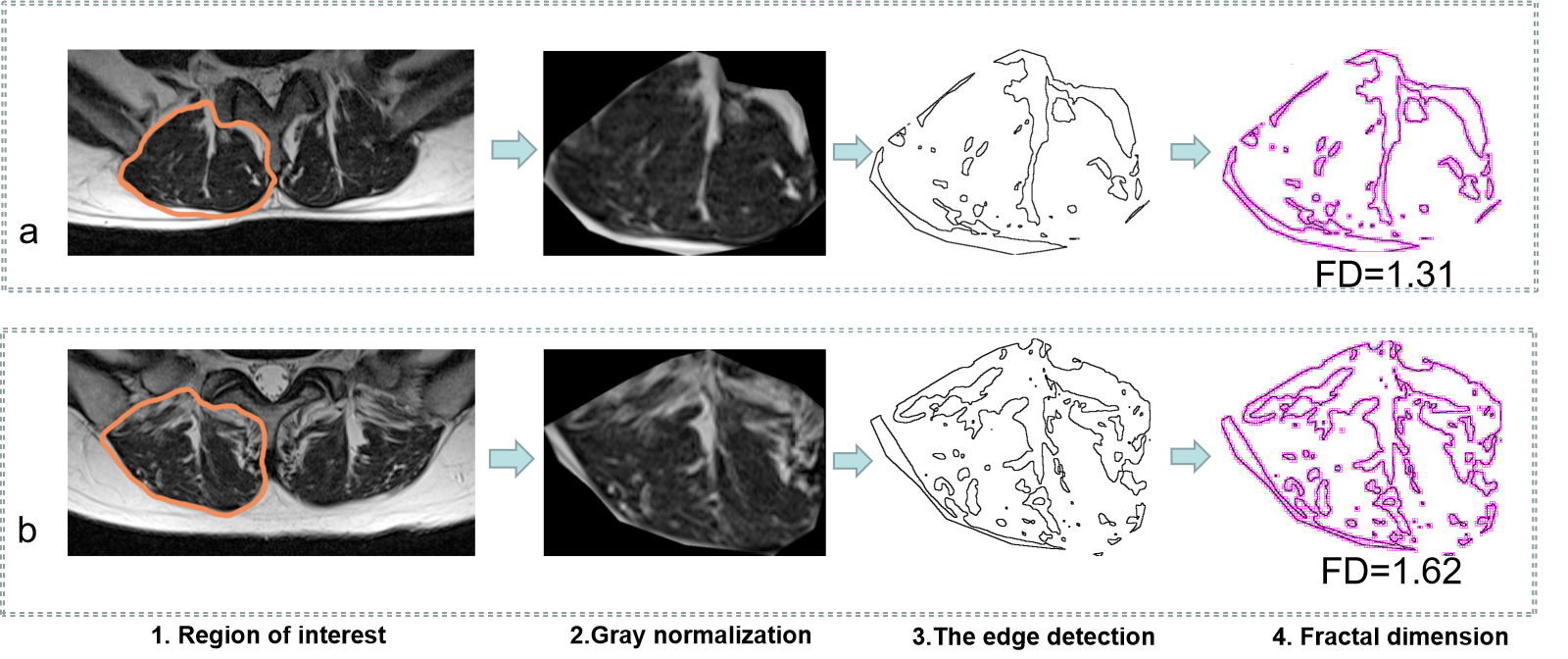
A population-based study collected MRI scans of about 213 adult subjects (18-85 years old). The information of LBP was determined by questionnaires previously used in the Danish population. In addition, information on lifestyle factors is provided to make it possible to link this link to potential changes/confusion effects of gender, obesity, type of work and physical activity. The lumbar multifidus muscle was analyzed using axial T2WI images. Data were acquired using a 3.0 Tesla MRI scanner. The sequence parameters of axial T2WI are as follows: repetition time 3500 m/s, echo time 90 m/s, 256×256 matrix, section thickness 4 mm, intersection gap 0.5 mm. Fractal analysis was performed using the ImageJ software (Wayne Rasband, National Institutes of Health, Bethesda) together with the FracLac plug-in (A. Karperien – Charles Sturt University, Australia). Fractal dimension were calculated accordingly to the box counting algorithm as the slope of the regression line for the log-log plot of the scanning box size and the count from a box-counting scan (Figure 2). Multiple linear regression analysis was used to explore the associations between low back pain and fractal dimension of multifidus muscle.Result
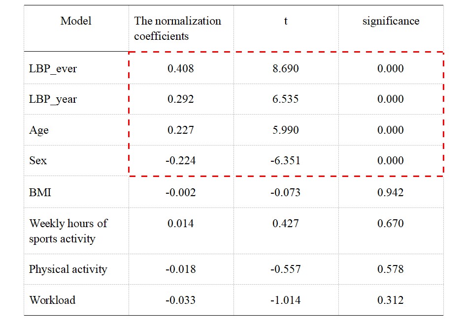
Multiple linear regression analysis showed that LBP-ever, LBP-year, age and sex had significant associations with fractal dimension of multifidus muscle. LBP-ever had the highest standardization coefficient (0.408, P<0.01), followed by LBP-year (0.292, P <0.01). Age and sex also had significant effects on the fractal dimension of multifidus muscle, but the effect was relatively low (0.227, P < 0.01; -0.224, P<0.01). The normalization coefficients and significance levels of all variables are shown in Table 1. The fractal dimension of female subjects was higher than that of male subjects. The average FD of female subjects was about 1.53, and that of male subjects was 1.36.Discussion and conclusion
It is found that the fractal dimension of LMM is closely related to LBP. The association was not affected by BMI, type of work or level of physical activity during leisure. Patients with low back pain had higher fractal dimension of multifidus muscle, although the main direction of this association remains unclear. Does lack of muscle strength or control lead to low back pain, or does low back pain affect muscles and their functions? However, the results of this study can show that it is feasible to decode the degree of multifidus degeneration by fractal dimension. The reason why low back pain is significantly related to fractal dimension may be that the fractal analysis method can effectively reflect signal heterogeneity of multifidus muscle, and the factors causing signal heterogeneity (fat infiltration, tear, etc.) have been reported to be independently related to pain3. In conclusion, the fractal dimension of LMM may be a reliable imaging marker for monitoring the degree of multifidus muscle degeneration in symptomatic patients.Acknowledgements
References
[1] Kjaer P, Bendix T, Sorensen J S, et al. Are MRI-defined fat infiltrations in the multifidus muscles associated with low back pain?. BMC Med. 2007, 15(5): 2-6.
[2] Gumussoy I, Miloglu O, Cankaya E, et al. Fractal properties of the trabecular pattern of the mandible in chronic renal failure. Dentomaxillofac Radiol. 2016,45(5): 20150389.
[3] Hildebrandt M, Fankhauser G, Meichtry A, Luomajoki H. 2017. Correlation between lumbar dysfunction and fat infiltration in lumbar multifidus muscles in patients with low back pain. BMC Musculoskelet Disord.2017, 18(1): 12-15.
Figures