2699
Stiffness change of the rotator cuff muscle before and after the tendon tear with magnetic resonance elastography and ultrasound elastography.
Akihisa Koga1, Yoshiaki Itoigawa1, Mikio Suga2, Yuri Suganuma3, Tomoki Wada1, Daichi Morikawa1, Yuichiro Maruyama1, and Kazuo Kaneko4
1Orthopaedic surgery, Juntendo Urayasu Hospital, Chiba, Japan, 2Center for Frontier Medical Engineering, Chiba University, Chiba, Japan, 3Graduate School of Science and Engineering, Chiba University, Chiba, Japan, 4Orthopaedic surgery, Juntendo University, Tokyo, Japan
1Orthopaedic surgery, Juntendo Urayasu Hospital, Chiba, Japan, 2Center for Frontier Medical Engineering, Chiba University, Chiba, Japan, 3Graduate School of Science and Engineering, Chiba University, Chiba, Japan, 4Orthopaedic surgery, Juntendo University, Tokyo, Japan
Synopsis
The purpose of this study was to explore the feasibility of Magnetic Resonance Elastography (MRE) measurement for quantification of the stiffness change of the shoulder rotator cuff muscle compared with Shear Wave ultrasound Elastography (SWE). Six porcine shoulders were used in this study. MRE and SWE measurement of the rotator cuff muscle was performed before and after the rotator cuff tendon detachment. Stiffness values were significantly lower after the tendon detachment in both MRE and SWE measurements (p<0.05). This result suggests that MRE could be a feasible method for quantification of the rotator cuff muscle stiffness as well as SWE.
Purpose
Shoulder rotator cuff tear is present 25-50%,1 and one of the most common cause of the shoulder pain and disability. Arthroscopic surgical repair is performed when conservative treatment for symptomatic rotator cuff tear has failed, however re-tear rate is remaining relatively high. Previous studies have reported re-tear rates are estimated to occur 17 to 70% of cases.2,3,4 To repair the rotator cuff tendon with the poor muscle extensibility, large tensile force is required, and excessive stress leads the re-tear. From this reason, it is important to predict the extensibility of the rotator cuff muscle for rotator cuff repair. Shear wave ultrasound elastography (SWE) has been used to measure stiffness of the supraspinatus muscle, which is the one of the shoulder rotator cuff muscles, in resent several studies.5,6 However, SWE has some disadvantages specific to the ultrasound imaging. The measurement was dependent on each measurer’s ability, and SWE could view only narrow space. On the other hand, magnetic resonance elastography (MRE) is the technique for measuring the stiffness using MRI.7 Previous studies reported that MRE was used to measure the properties of tissues; for example, liver, breast, brain and muscle.8-11 However, there have been few studies to investigate rotator cuff muscle stiffness using MRE. The purpose of this study was to explore the feasibility of MRE measurement for quantification of the supraspinatus muscle stiffness compared with SWE.Methods
Six porcine shoulders were used in this study. The subcutaneous soft tissues except for rotator cuff muscle were removed. Humeral bone was fixed 0˚ abduction position with the wire. SWE measurements were performed with an ACUSON S2000 (Siemens, Germany). The probe was placed on the center of the supraspinatus muscle surface and parallel to the muscle fiber. At the depth of 1cm from surface, we measured the shear wave propagation speed and calculated the stiffness. MRE measurement was performed with 0.3 T open MRI (Hitachi, Japan). 62.5 Hz vibration was propagated from a pneumatic driver system to create shear wave in the supraspinatus muscle. A motion encoding gradient (MEG) was added to gradient echo sequence to acquirer shear wave images of the tissue. The stiffness was estimated from the wave images using local frequency estimation (LFE) methods.12 The passive driver was placed on the center of the supraspinatus muscle surface. After the first measurements, supraspinatus and infraspinatus tendons were detached from the humeral bone. Then, MRE and SWE measurements were performed using the same manner again. A paired T-test was used to evaluate the difference of the results between before and after the detachment of the tendon.Result
Figue.1 shows the result of the MRE measurement. The images in the middle side are the wave images which indicate the tissue displacements due to vibrations. The region of the interest was set on the center of the supraspinatus muscle, and the mean stiffness was measured. In the MRE measurement, the mean stiffness value of the supraspinatus muscles in six shoulders were 12.6 ± 3.2 kPa before the tendon detachment, and 9.9 ± 2.7 kPa after the detachment. In the SWE measurements, mean stiffness value was 10.2 ± 2.2 kPa before the tendon detachment, and 8.1 ± 1.4 kPa after the detachment (Fig 2). Stiffness values were significantly lower after the tendon detachment in both MRE and SWE measurements (p<0.05).Conclusion
There were some previous studies that the supraspinatus muscles could be measured using MRE.13,14 However, these studies used only healthy shoulders, and there had been no report that investigated the stiffness change of the rotator cuff muscle before and after the rotator cuff tendon tear. In this study, we made a model detached the rotator cuff tendon using the porcine shoulders and could measure the stiffness change of the supraspinatus muscles. Our result shows stiffness values of the supraspinatus muscle became lower just after the rotator cuff detachment in both MRE and SWE measurement. It seems that the supraspinatus muscle became softer because the muscle tonus was decreased. Our result also demonstrated that MRE measurement showed the same tendency as SWE before and after rotator cuff detachment. This result suggests that MRE could be a feasible method for quantification of the rotator cuff muscle stiffness as well as SWE.Acknowledgements
No acknowledgement found.References
- Yamamoto A, Takagishi K, Osawa T, et al. Prevalence and risk factors of a rotator cuff tear in the general population. J Shoulder Elbow Surg 2010;19(1):116-120.
- Bartl C, Kouloumentas P, Holzapfel K, et al. Long-term outcome and structural integrity following open repair of massive rotator cuff tears. Int J Shoulder Surg 2012;6(1):1-8.
- Iannotti JP, Deutsch A, Green A, et al. Time to failure after rotator cuff repair: a prospective imaging study. J Bone Joint Surg Am 2013;95(11):965-971.
- Kim JR, Cho YS, Ryu KJ, et al. Clinical and radiographic outcomes after arthroscopic repair of massive rotator cuff tears using a suture bridge technique: assessment of repair integrity on magnetic resonance imaging. Am J Sports Med 2012;40(4):786-793.
- Itoigawa Y, Sperling JW, Steinmann SP, et al. Feasibility assessment of shear wave elastography to rotator cuff muscle. Clin Anat 2015;28(2):213-218.
- Itoigawa Y, Maruyama Y, Kawasaki T, et al. Shear Wave Elastography Can Predict Passive Stiffness of Supraspinatus Musculotendinous Unit During Arthroscopic Rotator Cuff Repair for Presurgical Planning. Arthroscopy 2018;34(8):2276-2284.
- Muthupillai R, Lomas DJ, Rossman PJ, et al. Magnetic Resonance Elastography by Direct Visualization of Propagating Acoustic Strain Waves. Science, 269:1854-1857, 1995.
- Mariappan YK, Glaser KJ, Ehman RL. Magnetic resonance elastography: a review. Clin Anat 2010;23(5):497-511.
- Numano T, Mizuhara K, Hata J, et al. A simple method for MR elastography: a gradient-echo type multi-echo sequence. Magn Reson Imaging 2015;33(1):31-37.
- Green MA, Geng G, Qin E, et al. Measuring anisotropic muscle stiffness properties using elastography. NMR Biomed 2013;26(11):1387-1394.
- Debernard L, Robert L, Charleux F, et al Analysis of thigh muscle stiffness from childhood to adulthood using magnetic resonance elastography (MRE) technique. Clin Biomech (Bristol, Avon) 2011;26(8):836-840.
- Manduca A, Oliphant TE, Dresner MA, et al. Magnetic resonance elastography: non-invasive mapping of tissue elasticity. Med Image Anal 2001;5(4):237-254.
- Ito D, Numano T, Mizuhara K, et al. A new technique for MR elastography of the supraspinatus muscle: A gradient-echo type multi-echo sequence. Magn Reson Imaging 2016;34(8):1181-1188.
- Hong SH, Hong SJ, Yoon JS, et al. Magnetic resonance elastography (MRE) for measurement of muscle stiffness of the shoulder: feasibility with a 3 T MRI system. Acta Radiol 2016;57(9):1099-1106.
Figures
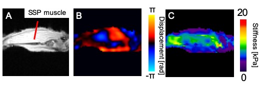
(A) An axial magnitude image of the supraspinatus muscle after the rotator cuff tendon detachment. (B) A wave image, which indicates the amplitude of the propagating shear wave. (C) A stiffness map obtained from the wave image.
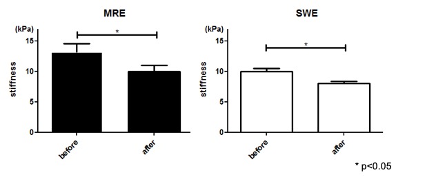
The mean stiffness values of MRE and SWE measurements before and after rotator cuff detachment. The stiffness values were significantly lower after the tendon detachment in both MRE and SWE measurements (p<0.05)