2691
Classification of annular fissures and pain-positive discograms using multiple MRI-features with Machine Learning1Institute of Clinical Sciences, Gothenburg University, Gothenburg, Sweden, 2Medical Physics and Biomedical Engineering, Sahlgrenska University Hospital, Gothenburg, Sweden, 3Radiology, Sahlgrenska University Hospital, Gothenburg, Sweden, 4Orthopaedics, Sahlgrenska University Hospital, Gothenburg, Sweden
Synopsis
Imaging-based features are needed to improve the characterization of degenerative IVD-changes and possibility of finding a linkage between features and pain.
Multiple T2w-imaging-features and Machine-Learning was used for classification of fissures involving outer annulus and for pain-positive discograms.
Fissures were classified with high accuracy/precision using regional/heterogeneity features with/without axial loading of the spine. For pain-positive discograms, a larger number of such MRI-features contributed to the classification.
Findings suggest that multiple MRI-features, extracted from T2w-imaging, improve the classifications, and that regional/heterogeneity features extracted with both conventional imaging with the spine unloaded and with axial loading of the spine are of importance.
Introduction
To date, several studies have shown the value of quantitative MRI and axial loading during MRI (alMRI) for detection of degeneration-related IVD-changes using single parametric features, e.g. mean IVD T2-value measured with conventional MRI in the supine position (uMRI) or the separation between histogram peaks representing nucleus pulposus and annular fibrosis. However, these imaging features neither correlate with clinical symptoms, nor do they consistently predict the development of low back pain (LBP).1,2In this study, we examine the value of multiple MRI-features using Machine Learning techniques for improved characterization of degenerative IVD-changes, but also for the possibility of finding a linkage between MRI-features and pain.
Methods
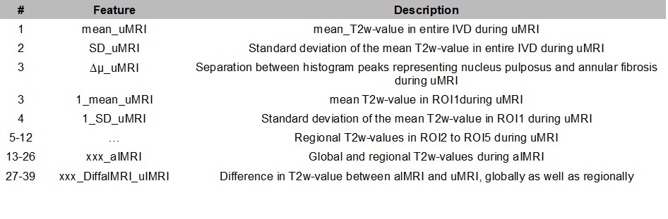
During one single day, MRI, CT and low-pressure discography (<50psi) were performed on the lumbar spine of each 30 LBP-patients. Quantitative MRI-features, which previously have been shown to correlate with IVD degeneration, were extracted from sagittal T2-weighted (T2w) images, acquired on a 1.5T system (Siemens Magnetom Symphony Maestro Class, Erlangen, Germany) using both uMRI and alMRI (TR=4ms,TE=124ms,slice thickness=4mm). Axial load was created with a compression device (DynaWell, Dynawell diagnostics AB, Las Vegas, NV USA) configured to generate axial compression of the lumbar spine, corresponding to half the patient's body weight.For each IVD, global and regional MRI-features, as well as heterogeneity features were extracted (Table 1) using an in-house segmentation program based on MatLab (R2018b, Mathworks, Natick, Massachusetts, U.S.A.). Global features were extracted from the entire IVD using regions of interest (ROIs) that were drawn semi-automatically on the 5 central slices of the T2w-images. Regional features were extracted from volumetric IVDs, dividing the ROIs into 5 equally large parts in the sagittal direction (ROI1 anterior to ROI5 posterior). The standard deviation of the mean and the difference between histogram peaks representing the nucleus pulposus and annular fibrosis were used as measures of IVD-heterogeneity.
Discograms were performed in 84 IVDs and labeled as pain-positive for concordant pain response at a pressure <50psi. Also, all CT-discograms were labeled by the reader according to the Dallas Discogram Description (DDD) and Adams grading, and then digitomized into annular fissures involving the outer annulus or not, i.e. DDD≥2/DDD≤1 respectively Adams≥D/Adams≤C.
The multiparametric analysis was performed using Anaconda with the Python distribution (Anaconda Software Distribution. Computer software. Vers. 2-2.4.0. Anaconda, Nov. 2016). Supervised “Decision Tree” Machine Learning was used to examine how well the included MRI-features classified the “true labels” DDD≥2/DDD≤1 and Adams≥D/Adams≤C, as well as pain-positive discograms. A 10-fold cross validation was used for model derivation and the supervised classifiers was applied to the remaining third of data in a separate data file containing unseen data. All data was normalized for quantification.
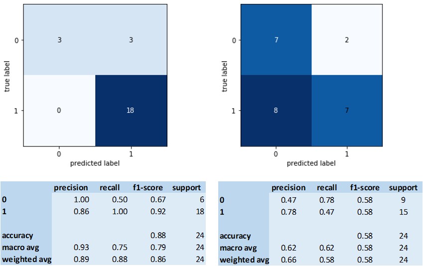
Results are presented as Decision Trees, confusion matrices and classification reports in terms of confusion matrix, accuracy, precision, recall, and F1 score (harmonic mean of the precision and recall).
Results
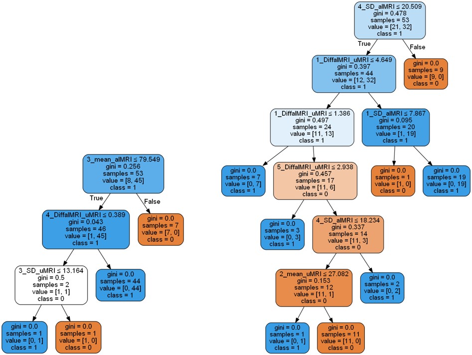
Figure 1 shows the evaluation of the “Decision Tree” algorithm for classification of fissures involving outer annulus (same results for DDD and Adams) and pain-positive discograms. Corresponding “Decision Trees” are shown in Figure 2. As can be seen in the figures, the algorithm classified IVDs with fissures involving the outer annulus with high accuracy and precision using only a few regional MRI-features, while a large number of regional and heterogeneity features were needed for the classification of pain-positive discograms, still, resulting in a lower accuracy and precision.Results from the cross validations showed high reproducibility for the fissure classification, mean:0.9 (std:0.1) and lower reproducibility for pain-positive discograms, mean:0.7 (std:0.1).
Discussion
This study suggests that multiple MRI-features, extracted from T2w-images, improve the classification of annular fissures and pain-positive discograms, and that regional as well as heterogeneity features using both uMRI and alMRI are of importance for the classifications.Especially, MRI-features that reflect IVD-behaviors during alMRI and alMRI-uMRI at the position of the nucleus pulposus (ROI3) and the transition zone between the nuclear pulposus and annular fibrosis (ROI4) were shown to be important for the fissure classification. Moreover, the heterogeneity of the IVD during uMRI at the position of the nucleus pulposus added a small contribution to the classification. The importance of adding image information from the central regions of the IVD for fissure classification may be explained by the associated remodulation of the nucleus pulposus.3,4
Regional and heterogeneity information seemed to be of importance also for the classification of pain-positive discograms. Especially, the heterogeneity of the IVD during alMRI at ROI4 and the load induced IVD-change at the most anterior and posterior sub-volume, i.e. ROI1 and ROI5, contributed the most to the pain classification. Thus, pain-positive discograms seem to be characterized by a significant loading effect on the annular fibrosis, but more importantly, by the variation in the IVD matrix structure at the transition zone, enhanced by alMRI.
Conclusion
This study demonstrates that multiparametric MRI improves the characterization of IVD fissures involving the outer annulus and pain-positive discograms and, thus, have an important value in future LBP-research. To enable direct clinical usability, additional features linked to pain needs to be added in order to improve detection of pain-positive spinal segments.Acknowledgements
No acknowledgement found.References
1. Doniselli et al. Eur Spine J 2018;27(11):2781-2790
2. Vagaska et al. Medicine (Baltimore) 2019;98(17):e15377.
3. Waldenberg et al. PLOS ONE 14(8)
4. Sharma et al. Spine (Phila Pa 1976). 2011;36(21):1794–800
Figures