2690
Accelerated high-resolution (0.5mm2) T1ρ mapping of the knee joint using interleaved spin-lock-prepared 3D gradient echo with Compressed SENSE1Saitama Medical University, Saitama, Japan, 2Philips Japan, Tokyo, Japan
Synopsis
3D T1ρ mapping sequence allows rapid acquisition of the entire volume data of each anatomic region, including major joint tissues. In this work, we propose the accelerated high-resolution 3D T1ρ-mapping of the knee-joint using motion-robust interleaved spin-lock acquisition with Compressed SENSE. We compared the T1p mapping with in-plane resolution of 0.5mm2 with different C-SENSE reduction factors (3 and 4.2). There are no clear differences between two image datasets, hence we applied C-SENSE reduction factor of 4.2 (5min48s) for clinical scans, acquiring motion-insensitive high-quality isotropic images less than 6 minutes.
Introduction
Spin-lattice relaxation in the rotating frame (T1ρ) has been shown to be sensitive to biochemical changes such as proteoglycan content of articular cartilage1. 3D T1ρ-weighted technique has been proposed to be a useful method allowing the rapid acquisition of the entire anatomic region2. Compared with 2D methods, 3D imaging is free from artifacts caused by slice cross-talk. Therefore 3D sequences can generally have a thinner slice thickness, and consequently may provide a more accurate assessment of cartilage degeneration. High-resolution MRI is particularly useful in the context of OA, in which cartilage becomes very thin—on the order of or less than 1 mm. Furthermore, a 3D acquisition is favorable due to the non-slice-selective nature of the T1ρ preparation pulses (spin-lock pulses).However, high-resolution 3D T1ρ-imaging was still challenging. One of the challenges was susceptibility to artifacts from inter-scan (or inter-shot) variability among respective spin-lock times, such as motion and physiological noise.
Recently, a unique interleaved spin-lock prepared steady-state free precession pulse sequence has been proposed to achieve single breath-hold T1ρ-mapping of the heart3, which employs different spin-lock pulses alternately before each gradient-echo train. Our strategy was to optimize and establish such interleaved spin-locking imaging for the knee-joint, which could be reducing inter-shot variability caused by motion or other factors related to quite long acquisition time.
On the other hand, a combination of parallel imaging and compressed sensing technique (Compressed SENSE, C-SENSE) has recently been developed to accelerate the acquisition time without increasing the image artifacts4,5. Accelerating the acquisition time can of course reduce the motion-related artifacts. Thus, the combination of both methods allows rapid and robust acquisition of 3D T1ρ mapping data. The purpose of this study was to demonstrate the clinical feasibility of accelerated high-resolution 3D T1ρ mapping of the knee-joint using motion-robust interleaved spin-lock acquisition with Compressed SENSE.
Methods
A total of seven subjects were examined on a 3.0T system (Ingenia Elition, Philips Healthcare). The study was approved by the local IRB, and written informed consent was obtained from all subjects. Schematic overview of the sequence for T1ρ-mapping we applied in this study is shown in Figure 1. T1ρ-mapping was performed using an inversion-recovery and T1ρ-prepared segmented gradient echo (turbo field-echo; TFE) sequence with water-selective excitation (ProSet). Inversion-recovery was used for suppression of synovial fluid. Five images with different SL-times (SL = 0, 10, 20, 30, 40ms) were acquired with interleaved acquisition. Amplitude of the SL pulse was set to 500 Hz. To optimize the scan protocol of 3D high-resolution T1ρ mapping in the knee-joint, we compared the T1ρ mapping with in-plane resolution of 0.5mm2 with different C-SENSE reduction factors. To validate the obtained T1ρ values, sagittal or coronal T1ρ maps were quantitatively compared. The average T1ρ values in the region-of-interests (ROIs) of the knee cartilage were used for comparison among respective spatial resolutions. We also compared with the T1ρ values reported in literature6. Imaging parameters for 3D high-resolution T1ρ mapping were: sagittal or coronal, TR=9.2ms, TE=4.7ms, ETL (TFE factor) =128, voxel size=0.5x0.5x3.0mm3, 25slices, TFE shot interval=5000ms, inversion delay=1760ms, ProSet1331, C-SENSE reduction factor=3 and 4.2, and total acquisition time=8min18s and 5min47s.Results and Discussion
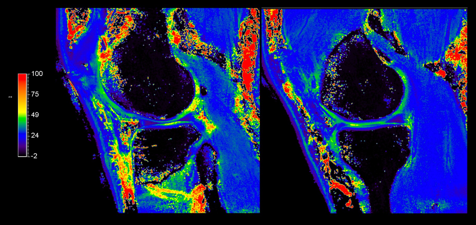
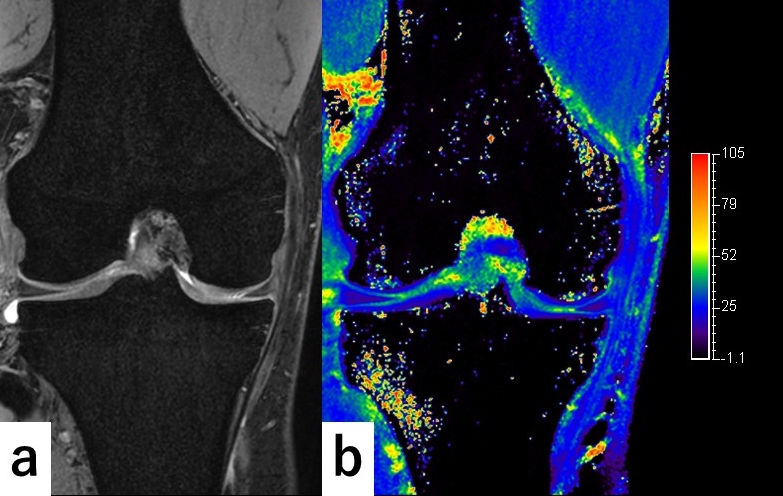
Figure 2 shows the comparison of two same spatial resolution 3D isotropic T1ρ-mapping with different Compressed SENSE (C-SENSE) reduction factors (3 with 8min18s, 4.2 for 5min48s). There are no clear differences between two image datasets, hence we applied C-SENSE reduction factor of 4.2 (5min48s) for clinical scans. Representative 3D high-resolution T1ρ-mapping with 0.5mm2 sagittal (Figure 3) and coronal (Figure 4) acquisition are shown. The optimized sequence provided motion-insensitive high-quality isotropic images less than 6 minutes. Figure 5b shows coronal 3D high-resolution T1ρ-mapping with 0.5mm2 in-plane resolution, which presents T1ρ-mapping of fine components of the knee, including articular cartilage, medial meniscus, medial collateral ligament, delineated by fat-saturated proton density weighted image (Figure 5a).Conclusion
The study demonstrated the clinical feasibility of accelerated 3D high-resolution (0.5mm2) T1ρ-mapping of the knee-joint less than 6 minutes using motion-robust interleaved spin-lock acquisition with C-SENSE. This technique might realize an rapid and robust acquisition of imaging data and allows an accurate assessment of cartilage degeneration and other fine components of the knee.Acknowledgements
References
1. Wáng, Y. X., et al. T1ρ magnetic resonance: basic physics principles and applications in knee and intervertebral disc imaging. Quantitative imaging in medicine and surgery. 2015 Dec; 5(6): 858–885.
2. Pakin SK, et al. 3D-T1ρ quantitation of patellar cartilage at 3.0T. J Magn Reson Imaging. 2006 Dec; 24(6): 1357-63.
3. van Oorschot JW, et al. Single Breath-Hold T1ρ-Mapping of the Heart for Endogenous Assessment of Myocardial Fibrosis. Invest Radiol. 2016 Aug; 51(8): 505-12.
4. Geerts-Ossevoort L, et al. Compressed SENSE Speed done right. Every time. The Netherlands: Philips Healthcare; 2018 Jan. Report No: 4522 991 31821. https://www.philips.de/content/dam/b2bhc/de/resourcecatalog/landingpages/ingeniaelition/White_Paper_Compressed_SENSE-opt.pdf
5. Mönch S, et al. Magnetic Resonance Imaging of the Brain Using Compressed Sensing - Quality Assessment in Daily Clinical Routine. Clin Neuroradiol. 2019 May 16. doi: 10.1007/s00062-019-00789-x. [Epub ahead of print]
6. Baboli R et al. Isotropic morphometry and multicomponent T1ρ mapping of human knee articular cartilage in vivo at 3T. J Magn Reson Imaging. 2018 Dec; 48(6): 1707-1716.Figures