2672
The value of MAVRIC-SL technique in evaluation of unicompartment knee arthroplasty (UKA) at 3.0TMR
Qiang Zhao1 and Lizhi Xie2
1Radiology Department, Peking University Third Hospital, Beijing, China, 2GE Healthcare, China, Beijing, China
1Radiology Department, Peking University Third Hospital, Beijing, China, 2GE Healthcare, China, Beijing, China
Synopsis
MAVRIC-SL is a new metal artifact suppression technology that mitigates this type of aliasing by combining the slice-selectivity of SEMAC with the overlapped excitation and combination properties of MAVRIC. In this study, 24 patients with the unicompartment knee arthroplasty (UKA) were performed MAVRIC-SL and conventional sequence scan at 3.0T MR. The effect of MAVRIC-SL on metal artifact subtraction was analyzed, the results show that MAVRIC-SL can significantly improve image quality and reduce image artifacts compared to conventional 2D-FSE.
Purpose
The subtractive effect of metal artifacts in the unicompartment knee arthroplasty (UKA) was compared between the MAVRIC-SL sequence and the conventional 2D-FSE sequence at 3.0T MR.Introduction
With the widespread of MRI, patients with metal implants in need of MR exams are not uncommon, such as post-operative evaluations[1]. Conventional MR scans are limited by the presence of metal artifacts which may lead to signal void, geometric distortion and signal pile-up due to severe field inhomogeneity[2]. MAVRIC-SL is a technique that may reduce metal artifact around small metal implants, both in-plane and through-plane artifacts may be reduced and hence improves visualization of the bone-implant interface, surrounding soft tissues, and influencing patient management directly[3,4]. In this study, the performance of MAVRIC-SL was investigated as compared to conventional 2D-FSE.Materials and Method
A total of 24 subjects with metal implants underwent MR exams on a 3.0T whole body scanner (GE discovery 750W, WI). Local ethical approval and consent forms were obtained. The MR protocol included both MAVRIC-SL and 2D-FSE. The overall image quality of the 24 groups of images was evaluated by two radiologists based on in plane artifacts and visibility of the implant and surrounding soft tissue image blurring. The difference between MAVRIC-SL and 2D-FSE was compared using the two-tailed Wilcoxon rank sum test and the paired Wilcoxon rank sum test was used to measure the artifact area of the comparison image and the changes in patient diagnosis caused by MAVRIC-SL imaging were recorded.Result
Representative FSE and MAVRIC-SL images of a patient with metal artifacts are shown in Figure 1 and 2. It was show that the use of MAVRIC-SL significantly reduced artifacts within and below the layer improves the discrimination between the implant interface and surrounding soft tissue and improves image quality (p<0.01); signicantly reduced image artifacts were observed on MAVRIC-SL (p<0.01); the MAVRIC-SL images feature higher level of blurring (p<0.01). Diagnosis based on MAVRIC-SL determined that 8 patients required surgery and surgical procedures and 13 patients did not require operation.Discussion and conclusion
Compared with conventional 2D-FSE sequences, MAVRIC-SL imaging of metal implant sites can significantly improve image quality and reduce image artifacts but with higher level of blurring.Acknowledgements
No acknowledgement found.References
1. Lee Y, Lim D, Kim E, et al. Feasibility of fat-saturated T2-weighted magnetic resonance imaging with slice encoding for metal artifact correction (SEMAC) at 3T. Magnetic Resonance Imaging. 2014; 32(8):1001–1005.2. Koch KM, Lorbiecki JE, Hinks RS, et al. A multispectral three-dimensional acquisition technique for imaging near metal implants. Magn Reson Med. Feb; 2009 61(2):381–390. [PubMed: 19165901]
3. Koch KM, Brau AC, Chen W, et al. Imaging near metal with a MAVRIC-SEMAC hybrid. Magn Reson Med. Jan; 2011 65(1):71–82.
4. Lu W, Pauly KB, Gold GE, et al. SEMAC: Slice Encoding for Metal Artifact Correction in MRI. Magn Reson Med. Jul; 2009 62(1):66–76.
Figures
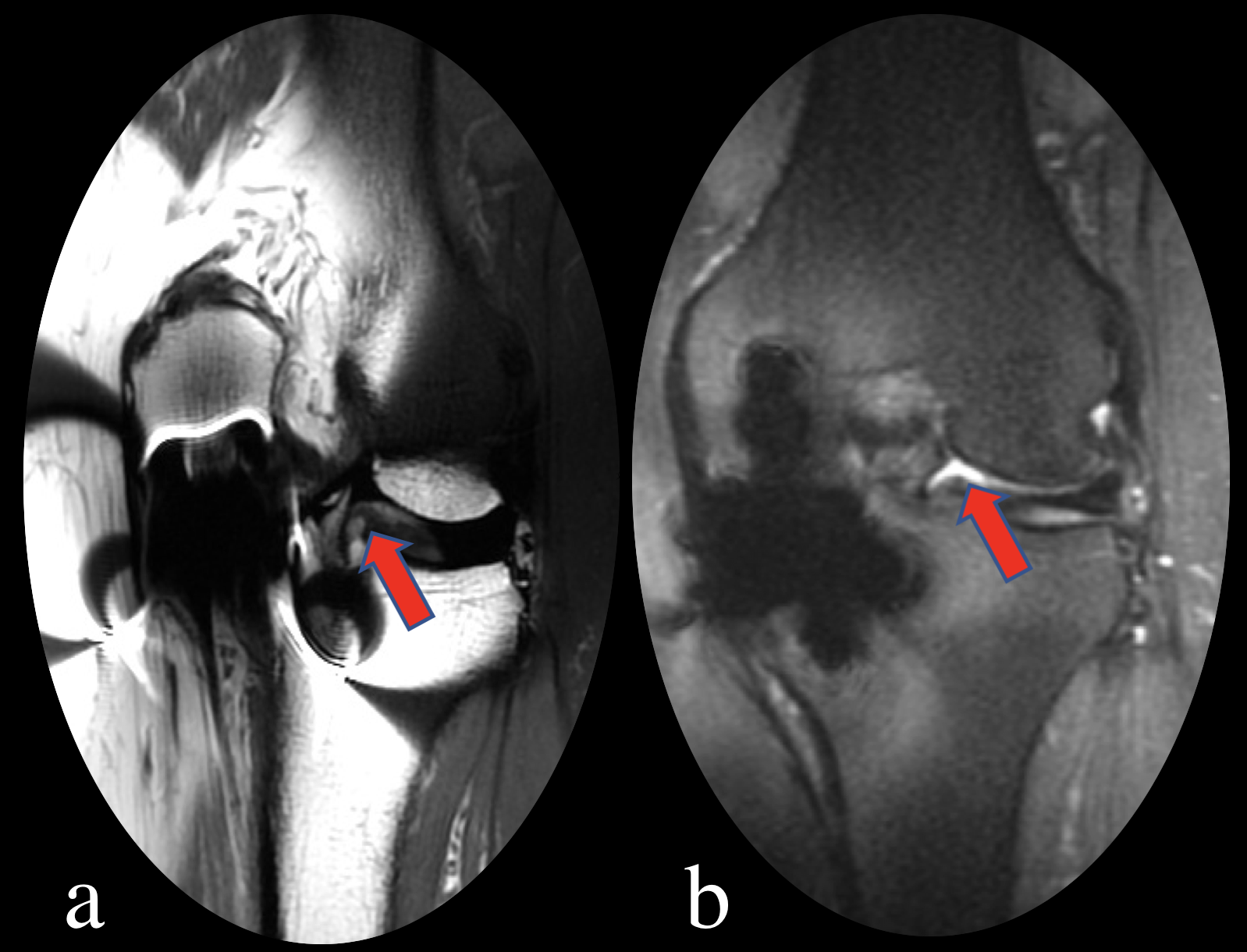
Figure.1 The same patient and the same level. (a) is a 2D-FSE fat suppression sequence scan
image; (b) is a MAVRIC-SL image, and the arrow indicates the synovial fluid
near the meniscus. (b) can be clearly seen than (a).
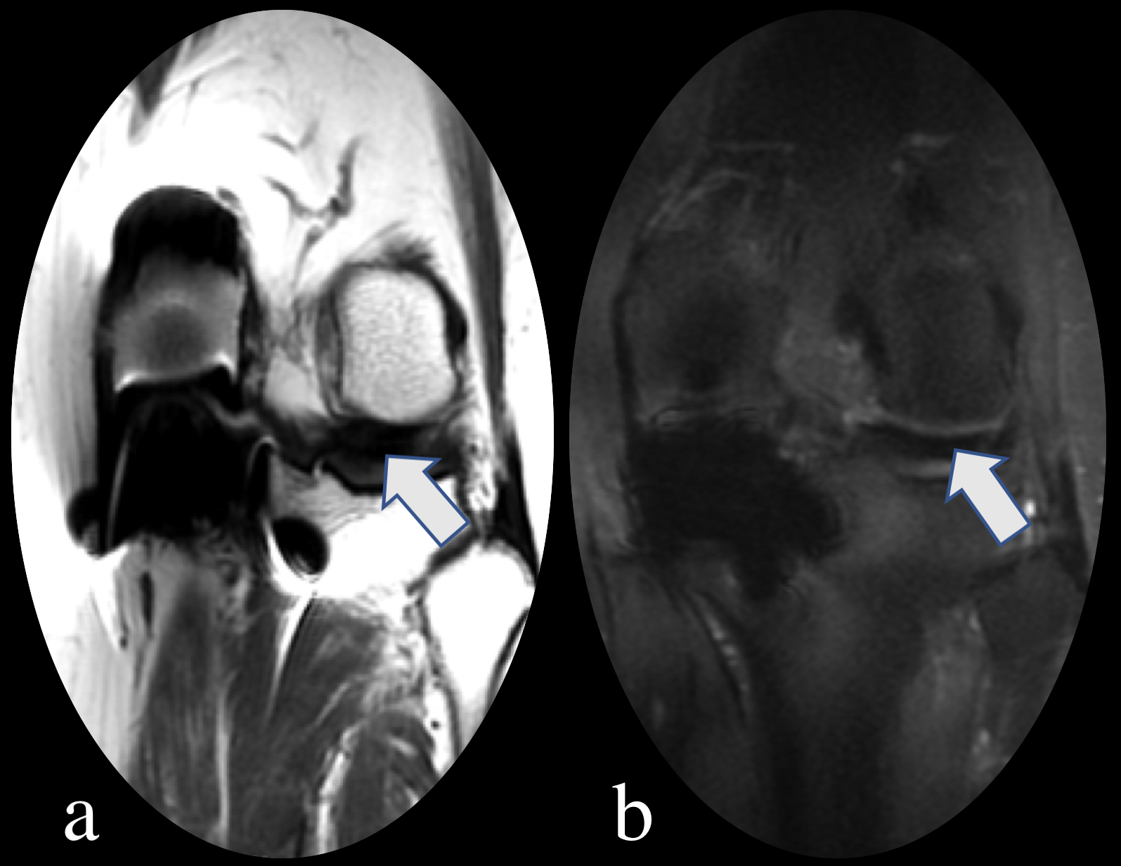
Figure.2 The arrow indicates that the contralateral
meniscus of the unicompartment knee arthroplasty. (a) shows the meniscus signal
is missing. (b) shows the contralateral meniscus more clearly.