2670
Value of texture analysis on the Ktrans map of dynamic contrast enhancement MRI for assessment of critical limb ischemia in diabetic rabbits1Renmin Hospital of Wuhan University, WuHan, China, 2GE Healthcare, Beijing, China
Synopsis
Critical limb ischemia (CLI) is a severe complication of diabetes which dramatically increases the risk of limb amputation and mortality. A greater degree of information may be derived by including an assessment of microvascular permeability in proximal femur by the transfer constant (Ktrans) values and MR texture analysis. Twelve diabetic rabbits (6 without CLI, 6 with occluded vasculature of the lower extremities) were examined by dynamic contrast‐enhanced magnetic resonance imaging at fixed time points.We found that Ktrans reached the minimum on one day after ischemia induction, and then recovered along with neoangiogenesis. Three texture features (mean value, MPP, sumAverage) were significantly different between two groups. Ktrans and three TA feature parameters were correlated with capillary density. Overall, Ktrans can be used to quantitative evaluation of changes in femur microvascular permeability in diabetic rabbits with CLI.. Texture analysis can provide more quantification information, which can be more accurately detecting alterations in bone marrow in diabetic rabbits
Critical limb ischemia (CLI) represents the leading cause of non–injury-related amputations and disabilities in diabetic patients. The prevalence rate is twofold in diabetic patients than in general population and leads to a higher risk of cardiovascular diseases1. MR texture analysis is an objective approach to quantify tissue gray-level patterns in provision of computer-assisted measurements that are independent of subjective visual interpretation2. But, to our knowledge, no studies have performed in vivo MR texture analysis on the bone marrow to assess CLI in diabetic patients. To monitor femur bone marrow injury and repair after femoral artery ligation in diabetic rabbits, the transfer constant (Ktrans) values were measured based on dynamic contrast-enhanced magnetic resonance imaging (DCE-MRI). The aim of our work is to assess the potential role of texture analysis (TA) for predicting critical limb ischemia in diabetic rabbits.
Materials and Methods
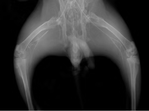
All experimental procedures were approved by the committee of the local institutional animal care and use. Twelve diabetic rabbits were randomly assigned to the critical limb ischemia group (n=6) or control (n=6) group .Critical limb ischemia in six rabbits was induced via unilateral ligation of the femoral artery. All rabbits underwent dynamic contrast-enhanced MR imaging examinations at fixed time points (1, 5, 10,15,20 and 25 days after surgery). Differences between the ligated and non-ligated limbs were assessed with independent-samples t-test, and analysis of variance was used to determine disease progression. Regions of interests in the proximal femur on Ktrans imaging were delineated to obtain 67 texture features. These features were compared between the two groups with an independent-samples t-test. Correlations of parameters calculated from DCE MR imaging (the Ktrans value and TA feature parameters) and microvessel density (MVD) were analyzed with the Pearson correlation coefficient. Receiver operating characteristic (ROC) curve and the area under the curve (AUC) analysis was applied to assess the discriminatory power of texture features. All statistical analyses were performed with SPSS 21.0.
Results and Discussion
All MR measurements of the ligated limb changed over time (p < 0.05), and disease progression in the ligated and non-ligated limbs differed significantly (p < 0.05). Ktrans reached the minimum on one day after ischemia induction, and then recovered along with neoangiogenesis, but the value maintained still but lower than the control group at post-ischemia 25 day.Luo et al3 has shown that DCE-MRI with Gd‐DTPA is useful to assess time‐dependent changes in muscle perfusion and perfusion reservation in the hind‐limb ischemia model. However, to date, no study has described the temporal changes in bone permeability in diabetic subjects with CLI via DCE MRI .
Three texture features (mean value, MPP, sumAverage) were significantly different between two groups. MVD was positively correlated with Ktrans and three TA feature parameters (p < 0.05 ). The area under the receiver operating characteristic curve was 0.985 for mean value, 0.985 for MPP , and 0.954 for sum of average.Mean value is the average of signal intensity of all pixels in each ROI. The mean value of positive pixels (MPP) reflected the average brightness of positive values in the images. On enhanced MRI images, blood vessels appear bright while ischemia areas may exhibit signal attenuation. Studies have shown that MPP has been correlated with angiogenesis in NSCLC4.
Conclusions
The transfer constant (Ktrans) value can be used to quantitative evaluation of changes in femur microvascular permeability in diabetic rabbits with chronic critical limb ischemia. DCE-MRI texture analysis of femur bone marrow may allow to detect alterations in bone marrow in diabetic rabbits. TA based on DCE-MRI could act as an imaging discriminator of critical limb ischemia in diabetes mellitus and serve as a new radiological analysis tool for monitoring diabetics progression.
Acknowledgements
No acknowledgement found.References
1. Iacopi E, Coppelli A, Riitano N, et al. Adherence to guideline recommended medical therapies in type 2 diabetic patients with chronic critical limb ischemia[J]. Diabetes Res Clin Pract,2019:107898.
2.Gillies R J, Kinahan P E, Hricak H. Radiomics: Images Are More than Pictures, They Are Data[J]. Radiology,2016,278(2):563-577.
3. Luo Y, Mohning K M, Hradil V P, et al. Evaluation of tissue perfusion in a rat model of hind-limb muscle ischemia using dynamic contrast-enhanced magnetic resonance imaging[J]. J Magn Reson Imaging,2002,16(3):277-283.
4.Ganeshan B, Goh V, Mandeville H C, et al. Non-small cell lung cancer: histopathologic correlates for texture parameters at CT[J]. Radiology,2013,266(1):326-336.
Figures