2651
Discrimination of subjects with and without knee osteoarthritis using a radiomics approach on tibial bone1Department of Radiology & Nuclear Medicine, Erasmus MC, Rotterdam, Netherlands, 2Department of Radiology & Nuclear Medicine and Department of Medical Informatics, Erasmus MC, Rotterdam, Netherlands, 3Department of General Practice, Erasmus MC, Rotterdam, Netherlands
Synopsis
Radiomic features were automatically extracted from tibial bone using knee MRI data of 665 women and the ability of the features to discriminate subjects with and without osteoarthritis was assessed. An area under the receiver operating characteristics curve of 0.80 was obtained for classifying subjects with and without osteoarthritis using an elastic net regression model that included radiomic features and covariates. Our results indicate that tibial bone characteristics are different between subjects with and without knee osteoarthritis.
INTRODUCTION:
Radiomics has been successfully applied to different magnetic resonance imaging (MRI) data [1], but it has not yet been widely used for assessment of knee osteoarthritis on MRI. The aim of this study was to automatically extract radiomic features from tibial bone using knee MRI data of a large population-based cohort and to assess the ability of radiomic features to discriminate subjects with and without osteoarthritis.METHODS:
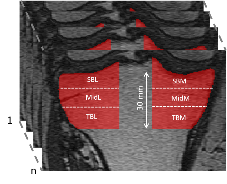
The study data consisted of 665 females from the Rotterdam Study scanned with a 1.5-T MRI scanner (Signa Excite 2, GE Healthcare). The mean (standard deviation) age and body mass index of the subjects were 54.6 (3.7) years and 26.8 (4.6) kg/m2, respectively. Right knees of the subjects and a fast imaging employing steady-state acquisition (FIESTA) sequence (repetition time: 5.6, time to echo: 1.8, flip angle: 35, voxel size: 0.3 x 0.3 x 1.2 mm3) was used in the quantitative analyses. The Medical Ethics Committee of Erasmus University Medical Center approved the study and all subjects provided a written consent. MRIs were scored according to the semi-quantitative MRI osteoarthritis knee score (MOAKS). Tibiofemoral osteoarthritis was defined as the presence of a full thickness cartilage loss and definite osteophyte, or one of the abovementioned features and two of the following features: partial-thickness cartilage loss, subchondral bone marrow lesion or cyst, or meniscal subluxation, maceration or degeneration [2].The tibial bone was segmented using an automatic segmentation method that combines multi-atlas and appearance models [3]. Twenty manually segmented tibias were used to train the segmentation method. The segmentation performance was evaluated on five manually segmented tibias. Altogether six 3-D volumes of interest (VOI) were automatically extracted from the medial and lateral compartments of the tibia (Figure 1). Radiomic features that are related to the shape and texture of the region, were calculated from each VOI using an open-source Workflow for Optimal Radiomics Classification (WORC) package in Python [1]. Features associated to the shape were calculated only for the whole tibial volume.
Elastic net, which is a regularized logistic regression method, was used for classification. We used 10-fold cross-validation with 100 repeats to train the models. Performance of the covariate (age and body mass index (BMI)) model, radiomic features model, and combined covariate + radiomic features model to discriminate subjects with and without osteoarthritis were assessed using an area under the receiver operating characteristics curve (ROC AUC). Statistical analyses and elastic net experiments were done using R software (version 3.5.2).
RESULTS:
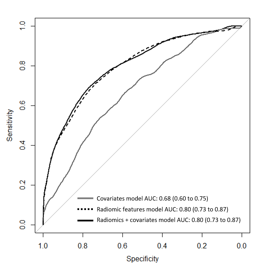
76 (11.5%) subjects had knee osteoarthritis. The mean (standard deviation) Dice score for the segmentation of the tibia was 0.96 (0.02). An ROC AUC value of 0.80 (95% confidence interval: 0.73 – 0.87) was obtained for classifying subjects with and without osteoarthritis using the model that included radiomic features and covariates (age and BMI) (Figure 2). When only radiomic features were used in the model, an ROC AUC of 0.80 (0.73 – 0.87) was obtained. Age and BMI alone yielded an ROC AUC of 0.68 (0.60 – 0.75).DISCUSSION:
We used an automatic method to extract radiomic features from tibial bone using a knee MRI data from a large population-based cohort. We found that tibial bone characteristics were different between subjects with and without knee osteoarthritis. The radiomic approach provides additional information on the bone changes in osteoarthritis.CONCLUSION:
Our results show that radiomic features of tibial bone on knee MRI are different between subjects with and without osteoarthritis.Acknowledgements
This project has received funding from the European Union’s Horizon 2020 research and innovation programme under the Marie Sklodowska Curie grant agreement No 707404.References
[1] Starmans et al., Classification of malignant and benign liver tumors using a radiomics approach, Medical Imaging 2018: Image Processing 10574, 105741D.
[2] Hunter et al., Definition of osteoarthritis on MRI: results of a Delphi exercise, Osteoarthritis and Cartilage 2011, 963-969.
[3] Hansson et al. Evaluation of two multi-atlas cartilage segmentation models for knee MRI: Data from the Osteoarthritis Initiative, IWOAI2016.
Figures