2645
Explore the performance of compressed-SENSE accelerated 3D mDIXON on kidney of healthy subjects1The First Affiliated Hospital of Dalian Medical University, Dalian, China, 2Philips Healthcare, Beijing, China
Synopsis
The compressed sensing (CS) technique, which implements k-space sparses ampling and iterative reconstruction algorithm, has now been widely used in acceleration of MR examinations of various anatomies and anatomical contrasts. This study aims to explore the performance of 3D water-fact imaging accelerated using a combination of compressed-sensing and sensitivity encoding on kidney of healthy subjects. Several acceleration factors of the proposed technique were compared against the traditional sensitivity-encoding technique and an acceleration factor of 2 was recommended for a balance between imaging time and image quality.
Purpose
To explore the performance of compressed-SENSE accelerated 3D mDIXON on kidney of healthy subjects.IntroductionIn
Recent years, the incidence of various kidney diseases has been increasing, and the The evaluation of kidney diseases has becoming increasingly important due to an increase in the incidence of various kidney diseases1.Due to its excellent soft tissue resolution, multi-parameter, multi-sequence and multi-orientation imaging features, MRI can clearly display the morphological manifestations of renal parenchyma and aggregation system, and can provide multiple diagnostic information for renal lesions 2.Due to the long scan time of MRI, its application scope is limited for patients who cannot hold their breath for a long time to perform visceral examination. Therefore, shortening the scan time without loss of image quality is still one of the active research fields for kidney MRI. Compressed SENSE (CS-SENSE) technology 3, a combination of compressed sensing 4 and sensitivity encoding, was proven to shorten the signal acquisition time5, However, there has been no research on the application of CS-SENSE on renal 3D water-fat imaging (mDIXON) sequence, which was the aim of this study.Materials and methods
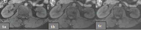
15 healthy volunteers (7 males, aged 24-65 (44.1±11.3) years, BMI=24.5±2.86) were recruited and under went the two-kidney 3D mDixon scans on a 3.0 T MR scanner(Ingenia CX, Philips Healthcare, Best, the Netherlands)using a 16-channel phase-arrayed coil (dS dorso, Philips Healthcare, Best, the Netherlands). The sequence was repeated 3 times for each volunteers, accelerated with SENSE factor of 2 (Group A), CS-SENSE factors of 2 (Group B) and 4 (Group C),respectively, scan durations 14.8, 12.9 and 7.2s.Two observers (5 and 3 years of abdominal MRI experience, respectively) performed subjective ratings and data quality measurements on the vendor-provided post-processing workstation(ISP).Criteria including identification of the medulla, image sharpness, image artifacts, and image noise were considered in subjective ratings. Measurements were performed to involve both kidney sat three levels, with ROIs were placed on the cortex(20 mm2)、medulla(30 mm2), and ipsilateral spinal muscles(30 mm2). Kidneys datas averaged respectively, and calculate the SNR and CNR, SNR = SI/SD muscles, CNRs were calculated between cortex and medulla. The intraclass correlation coefficient (ICC) was used to evaluate the consistency between measurements by the two observers. If the consistency is good, measurements of the senior observer would be tested for normality. Univariate analysis of variance (ANOVA) was used to check if the measurements conform to the normal distribution, and the LSD method was used for subsequent pair wise comparison. The Kruskal-Wallis test was used in the non-normal distributed measurements for inter-group comparison, and the different parameters were selected for subsequent pair-to-pair comparison.Results
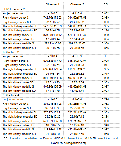
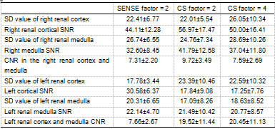
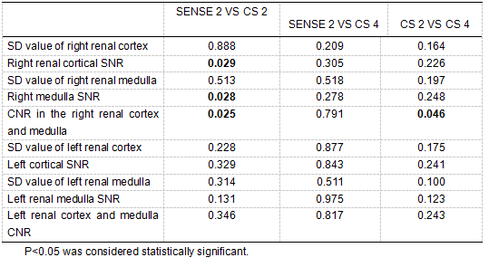
The obtained subjective scores, measure signal strengths (SI) and standard deviations (SD) for resulted images of the three groups were well consistent between the two observers (ICC > 0.75,Table 1).Images from the group B (CS factor = 2) are with the highest subjective scores (4.9±0.4), and with significantly higher image-quality values than group A (right cortical SNR, right medullary SNR, and right inter medullary CNR) and C (right intermedullary CNR). Other measurements of the right kidney and all measurements of the left kidney showed no statistical difference(Tables 2 and 3)Conclusion
In summary, the 3D mDIXON scan on kidney can be effectively accelerated by the CS-SENSE technique. In comparison with the traditional SENSE method, the CS-SENSE technique is associated with shorter scan time and better image quality. Based on the subjective ratings and image quality measurements, we recommend using the CS-SENSE technique with an acceleration factor of 2 for 3D water-fat imaging on kidney.Acknowledgements
No acknowledgement found.References
[1]Zebin Luo. imaging diagnosis of rare primary renal tumor . Imaging research and medical application.2019,3(16):217-218.
[2]HaoSun, Zhenhui Li, Zhengyu Jin. Research progress of magnetic resonance imaging in diagnosis of renal diseases . Radiology practice,2018,33(8):783-784.
[3]He M, Xu J, Sun Z, et al. Comparison and evaluation of the efficacy of compressed SENSE (CS) and gradient- and spin-echo (GRASE) in breath-hold (BH) magnetic resonance cholangiopancreatography (MRCP). J Magn Reson Imaging 2019; doi: 10.1002/jmri.26863
[4]Lusting M, Donoho D, Pauly JM. Sparse MRI: The application of compressed sensing for rapid MR imaging.Magn Reson Med, 2007;58(6):1182-1195.
[5] Jaspan ON, Fleysher R, Liption ML. Compressed sensing MRI: a review of the clinical literature. The British journal of radiology.2015,88(1056):20150487.
Figures