2642
Fat suppression efficiency in kidney diffusion imaging with different techniques
Jingjun Wu1, Ailian Liu1, Jiazheng Wang2, Zhongping Zhang2, Dahua Cui1, Lihua Chen1, Qingwei Song1, Renwang Pu1, and Bingbing Gao1
1Department of Radiology, the First Affiliated Hospital of Dalian Medical University, Dalian, China, 2Philips Healthcare, Beijing, China
1Department of Radiology, the First Affiliated Hospital of Dalian Medical University, Dalian, China, 2Philips Healthcare, Beijing, China
Synopsis
The reverse gradient technique showed equal fat suppression efficiency and higher SNR and CNR when compared to the RF inversion method in liver DWI, while the measured ADC values were consistent among the two. Chemical shift artifacts remain problematic in abdominal diffusion imaging due to the challenges in B0 field shimming, and the adoption of the optimal fat suppression technique for different application scenarios is important to balance between fat suppression, SNR, and ADC accuracy.
Purpose
To objectively compare the fat suppression efficiencies of two techniques for kidney diffusion weighted MRI (DWI) on image quality and apparent diffusion coefficients (ADC) quantification.Materials and Methods
Ten health subjects (mean age 28.7±2.15 years) were prospectively involved in the study and underwent 3.0T MR scan (Ingenia CX, Philips) with a 16-channel abdominal array coil. The MR protocols included reduced FOV DWI sequences (single diffusion encoding direction, b values = 0, 1000, 2000) with the same TR/TE (1394/75) but two different fat suppression techniques. In the first technique, the fat and water proton magnetization were both inverted using an adiabatic RF pulse and the acquisition started when the former relaxed to 0 in the longitudinal direction. In the second technique, the excitation and refocusing RF pulses experienced gradients with opposite polarities such that the fat slice was excluded from refocusing due to the chemical shift. All image analysis was performed on IntelliSpace Portal (Philips) (Figure 1). Two radiologists independently placed the region of intcrest (ROI) on renal cortex and medulla at upper, mid and lower pole of kidney levels, and on the psoas muscle of the same level, respectively, where the ADCs, SNRs, and CNRs (kidney to psoas muscle) were measured. These measurements between images from different sequences were analyzed using Friedman-test (SSPS, IBM).Results
The SNR and CNR at b = 1000/2000 in the renal cortex and medulla at mid pole in the technique 2 group were both significantly higher than those in the technique 1 group (P<0.05). There was no statistical difference in SNR and CNR in other regions between the two techniques. However, the mean SNR and CNR was significantly higher in technique 2 group (P<0.05) (Table 1 and 2). Notably, there was no statistical difference in ADC values between the two techniques (Table 3).Discussion and Conclusion
The reverse gradient technique showed equal fat suppression efficiency and higher SNR and CNR when compared to the RF inversion method in liver DWI, while the measured ADC values were consistent among the two. Chemical shift artifacts remain problematic in abdominal diffusion imaging due to the challenges in B0 field shimming, and the adoption of the optimal fat suppression technique for different application scenarios is important to balance between fat suppression, SNR, and ADC accuracy.Acknowledgements
No acknowledgement found.References
No reference found.Figures
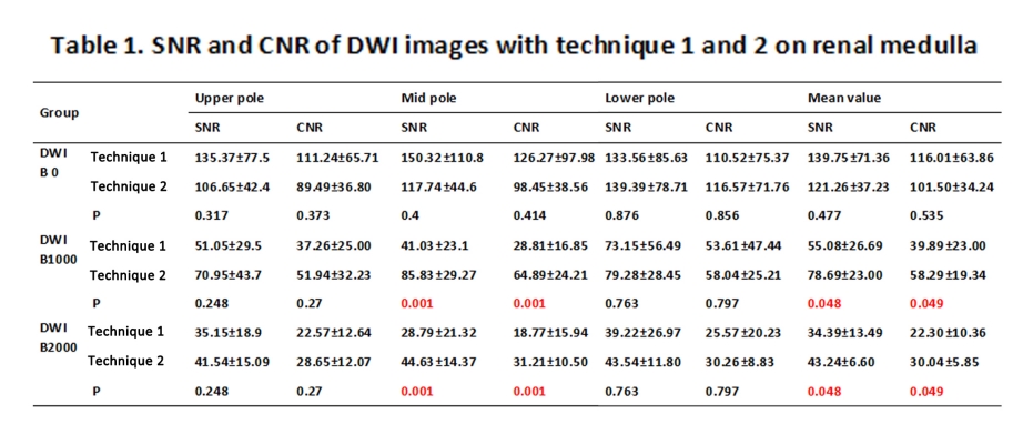
Table 1
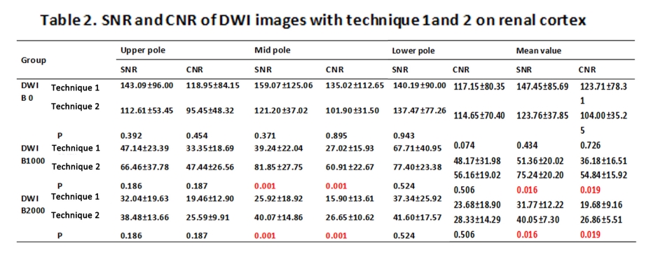
Table 2
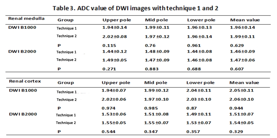
Table 3
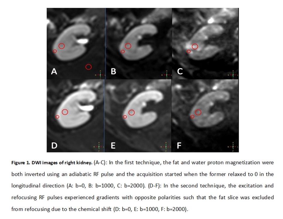
Figure 1