2630
A preliminary study on the value of amide proton transfer-weighted imaging in differentiating primary from secondary malignant liver tumors1The First Affiliated Hospital of Dalian Medical University, Dalian, China, 2Philips Healthcare, Beijing, China
Synopsis
Our work aimed to explore the value of amide proton transfer-weighted (APTw) imaging in diagnosing primary and secondary malignant liver tumors. The result manifested that the novel imaging tool had a valuable utility in differentiating primary from secondary malignant liver tumors (AUC: 0.879; sensitivity: 76.9%; specificity: 100%).
Introduction
Hepatocellular carcinoma (HCC) was one of the common fatal malignant tumors[1]. Compared with other anatomic sites, liver was the more commonly seen organ to which various primary malignant tumors metastasized[2,3]. Given that primary and secondary liver tuomrs had distinct treatment and prognosis, the differential diagnosis of both was critical. Amide proton transfer-weighted (APTw) imaging is a novel MRI imaging tool based on the endogenous substance in tissue, namely the amide protons in mobile cellular proteins and peptides[4]. Previous studies have shown that APTw imaging was applied to diagnose central nervous system diseases and cervical cancer[5,6]. Empirically, the application of APTw imaging in liver and related literature reports, especially the diagnosis of primary and secondary malignant liver tumors, are rare.Materials and Methods
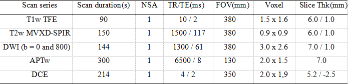
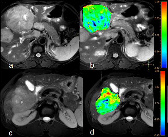
This study has been approved by the local IRB. Twenty cases of liver lesions based on clinical symptoms and image preliminary diagnosis in our hospital were retrospectively analyzed, including 13 cases of primary liver tumors (12 males and 1 female; (59.92±8.24) years old with 9 HCC and 4 intrahepatic cholangiocellular carcinoma and 7 cases of secondary liver tumors (3 males and 4 females; (59.50±7.33) years old). The T1-wighted (T1w) turbo field echo (TFE), T2-weighted (T2w) with motion correction technique MultiVane XD (MVXD) and Spectral Presaturation with Inversion Recovery (SPIR) for fat suppression, diffusion weighted imaging (DWI), APTw and dynamic contrast enhanced (DCE) sequences were performed on clinical whole-body 3.0-Testla MR system (Ingenia CX 3.0T, Philips Healthcare, Best, the Netherlands) with the scan parameters shown in Table 1. APTw signal intensity (SI) was defined as the asymmetry magnetization transfer ratio at 3.5 ppm and was mapped. Referring to the lesion information obtained from T2-weighted images, two radiologists manually placed circle ROIs (100-200 mm2) on the axial slice indicating the largest liver lesions on APTw images . The average APTw values were calculated to minimize measurement bias. The consistency of APTw measurements of lesions between the two radiologists were tested using intra-class correlation coefficients (ICC) in SPSS (IBM). APTw values were compared between two groups using Mann-Whitney U test. Receiver operating characteristic (ROC) curve was drawn to analyze the diagnostic efficiency of the parameter in differentiating the primary from secondary malignant liver tumors.Results
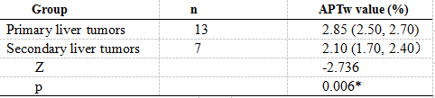
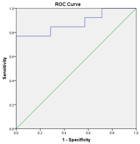
There was a good agreement between two observers with ICC = 0.934. APTw values of the primary liver tumors and the secondary liver tumors were 2.85(2.50,2.70) % and 2.10(1.70, 2.40)%, respectively, with a significant difference (p = 0.006) (Table 2). Area under the curve (AUC) for APTw values which is used to differentiate the primary liver tumors from secondary liver tumors was 0.879 and the feasible threshold was 2.625 with sensitivity of 76.9% and specificity of 100% (Figure 1).Discussion and Conclusion
Our study revealed that the primary liver tumors had significantly higher APTw values than secondary liver tumors, which was similar to the previous literature researching for solid brain metastases and glioblastomas[7].APTw imaging, as a novel technology generating image contrast without the contrast media, has a potential in the differential diagnosis of primary and secondary malignant liver tumors, and can provide a non-invasive approach for detecting primary and secondary malignant liver tumors.
Acknowledgements
No acknowledgement found.References
1. Motola-Kuba D, Zamora-Valdés D, Uribe M et al. Hepatocellular carcinoma. An overview. Ann Hepatol, 2006, 5 (1): 16-24.
2. Thomassen I, van Gestel YR, Lemmens VE et al. Incidence, prognosis, and treatment options for patients with synchronous peritoneal carcinomatosis and liver metastases from colorectal origin. Dis. Colon Rectum, 2013, 56 (12): 1373-80.
3. Rajasekaran Rathnakumar G, Gupta A, John F et al. A case of gastric small cell carcinoma with metastases to bone and liver. J Gastrointest Oncol, 2019, 10 (5): 1027-1031.
4. Zhou J, Heo HY, Knutsson L et al. APT-weighted MRI: Techniques, current neuro applications, and challenging issues. Magn Reson Imaging, 2019, 50 (2): 347-364.
5. Debnath A, Gupta RK, Singh A. Evaluating the Role of Amide Proton Transfer (APT)-Weighted Contrast, Optimized for Normalization and Region of Interest Selection, in Differentiation of Neoplastic and Infective Mass Lesions on 3T MRI. Mol Imaging Biol, 2019, undefined: undefined.
6. He YL, Li Y, Lin CY et al. Three-dimensional turbo-spin-echo amide proton transfer-weighted mri for cervical cancer: A preliminary study. Magn Reson Imaging, 2019, 50 (4): 1318-1325.
7. Yu H, Lou H, Zou T et al. Applying protein-based amide proton transfer MR imaging to distinguish solitary brain metastases from glioblastoma. Eur Radiol, 2017, 27 (11): 4516-4524.
Figures