2616
Diffusion-Weighted Imaging of the Liver with Stretched-Exponential Model
Takeshi Yoshikawa1, Yoshiharu Ohno2, Masao Yui3, Yoshimori Kassai3, Tatsuya Ohkubo3, Shinichiro Seki4, Katsusuke Kyotani5, and Yuji Kishida6
1Kobe University Graduate School of Medicine, Kobe, Japan, 2Fujita Health University School of Medicine, Toyoake, Japan, 3Canon Medical Systems Corporation, Otawara, Japan, 4Hyogo Prefectural Tamba Medical Center, Tamba, Japan, 5Kobe University Hospital, Kobe, Japan, 6Konan Medical Center, Kobe, Japan
1Kobe University Graduate School of Medicine, Kobe, Japan, 2Fujita Health University School of Medicine, Toyoake, Japan, 3Canon Medical Systems Corporation, Otawara, Japan, 4Hyogo Prefectural Tamba Medical Center, Tamba, Japan, 5Kobe University Hospital, Kobe, Japan, 6Konan Medical Center, Kobe, Japan
Synopsis
Stretched-exponential model enables diffusion analysis considering diffusion varieties in each voxel on abdominal DWI. Our study showed stretched-exponential model has a potential to increase diagnostic performance of liver DWI. Alpha has a potential to be used for malignant lesion differentiation.
Backgrounds & Purpose
Backgrounds- To measure water diffusion in tissue more precisely compared to conventional ADC (mono-exponential model), several non-mono-exponential models have been proposed and evaluated.
- One of them is stretched-exponential model.
- It can estimate heterogeneity of water diffusion as well as microstructures in the tissue and express them with only two parameters.
- Distributed diffusion coefficients (DDC)
- Water molecular diffusion heterogeneity index (Alpha, α)
α=0: high degree of multi-exponential signal decay
Equation : Sb/S0 = exp{-(b×DDC)α} cf. Sb/S0 = exp(-b×ADC)
Purpose
- The purpose of this study was to assess DWI with stretched-exponential model in evaluation of focal liver lesions
Methods and Materials
Patients & Lesion- 76 patients (53 men and 22 women, mean: 66.9 years), who were suspected to have hepatic malignancy and underwent 3T-MRI, were chosen and retrospectively analyzed.
- 78 malignant (HCC: 43, metastasis: 27, CCC: 3, combined HCC & CCC: 5) and 30 benign (hepatic cyst: 18, hepatic hemangioma: 12) lesions were confirmed and evaluated.
- All patients underwent MRI at a 3T scanner (Vantage Titan 3T; Canon Medical Systems, Otawara, Japan).
- Source DWIs were obtained with SE-EPI sequence (TR/TE/FA = 3000-6000/66/90, b values: 0, 500, 1000, matrix: 96 ×128, thickness: 7mm, NEX: 2, scan time: 10-12min, PASTA+SPAIR, PI: 2, MPG: x, y, z) as one of routine sequences in our institution.
- ADC, DDC, Alpha images were calculated by using mono-exponential and stretched-exponential models on a workstation (OleaSphere, Olea Medical).
- Oval ROIs were placed in the liver and focal lesions and mean ADC, DDC, and Alpha were measured.
- Mean values of malignant and benign lesions were compared for each parameter.
- Correlations coefficients among the three parameters were assessed in the liver and focal lesions.
- Lesion-to-liver contrasts (=(lesion-liver) / (lesion+liver)) were calculated and compared among the parameters.
- Lesion characterization was compared using ROC among the parameters and their combinations.
- Mean values of HCC, metastasis, and other primary cancers were compared for evaluation of lesion differentiation capability.
Results
- Malignant lesion ADC and DDC were significantly lower than benign ones (p<0.0001, fig. 1).
- DDCs showed the trends similar to ADCs in the liver and focal lesion and were significantly correlated. Correlations of lesion ADCs and DDCs were significantly lower than hepatic ones (fig. 2).
- Malignant lesion contrast was significantly higher on DDC than on ADC (p=0.019, fig. 3).
- For lesion characterization, no significant difference was found between ADC and DDC, and Az had non-significant trend toward highest at ADC+Alpha and ADC+DDC+Alpha sets (Az: 0.996, fig. 4).
- In malignant lesion differentiation, significant difference was found between HCC and metastasis on ADC (1.18 vs 1.39, p=0.009) and Alpha (0.71 vs 0.80, p=0.011), and between HCC and other primary cancers on Alpha (0.71 vs 0.92, p=0.0003) (fig. 5).
- Representative cases are shown in figures 6 & 7.
Summary & Conclusion
Summary- Stretched-exponential model enables diffusion analysis considering diffusion varieties in each voxel and has a potential to increase diagnostic performance of liver DWI.
- DDC can be used as an alternative to ADC.
- Alpha has a potential to be used for malignant lesion differentiation.
- Stretched-exponential model has a potential to increase diagnostic performance of liver DWI.
- Alpha has a potential to be used for malignant lesion differentiation.
Acknowledgements
No acknowledgement found.References
Previous reports for liver tumors and fibrosis
- Kim HC, et al. Eur Radiol 2019.
Alpha indicated degree of necrosis in treated colon ca metastasis.
- Hu Y, et al. Cancer Med 2018.
Alpha: malignant < benign lesion
- Seo N, et al. Eur Radiol 2018.
Alpha: F0-1 > F2 > F4
Figures
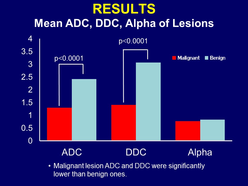
Figure 1. Mean
ADC, DDC,
Alpha of Lesions
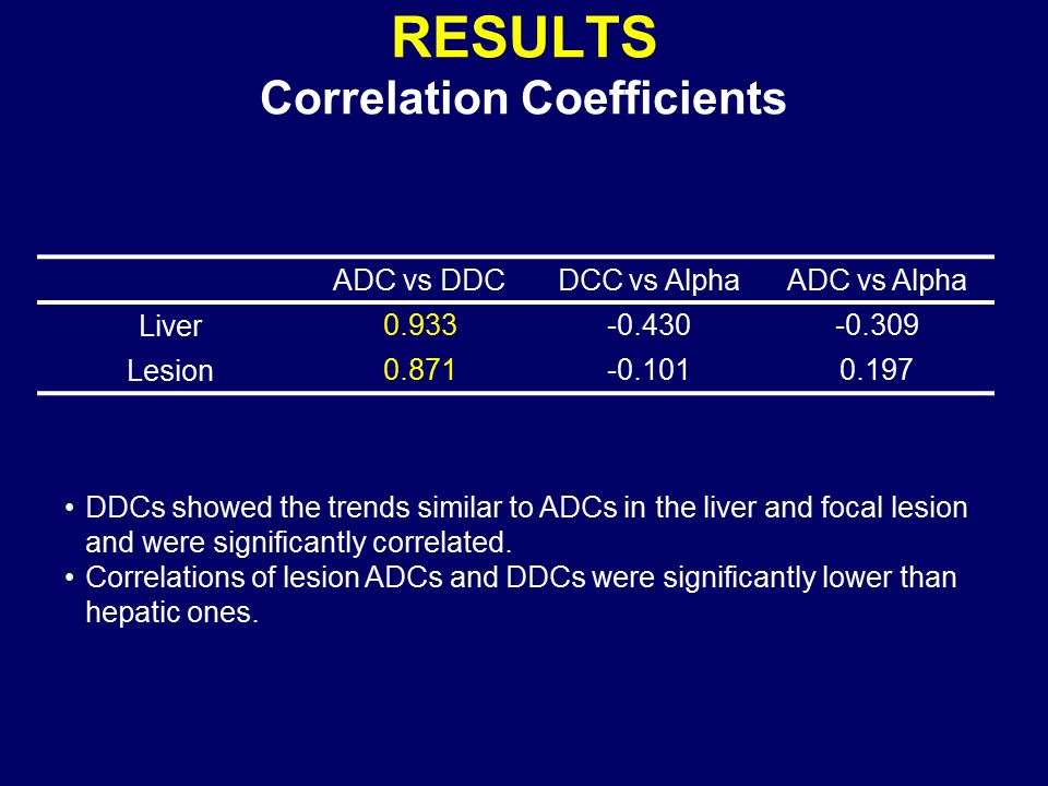
Figure 2. Correlation Coefficients
among the Parameters
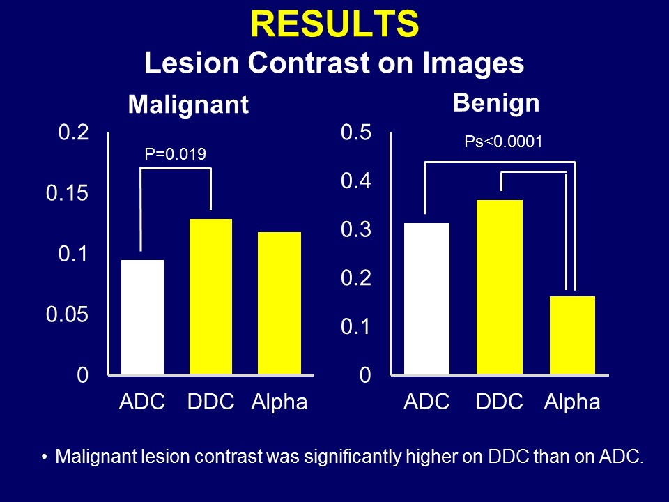
Figure 3. Lesion Contrast on Images
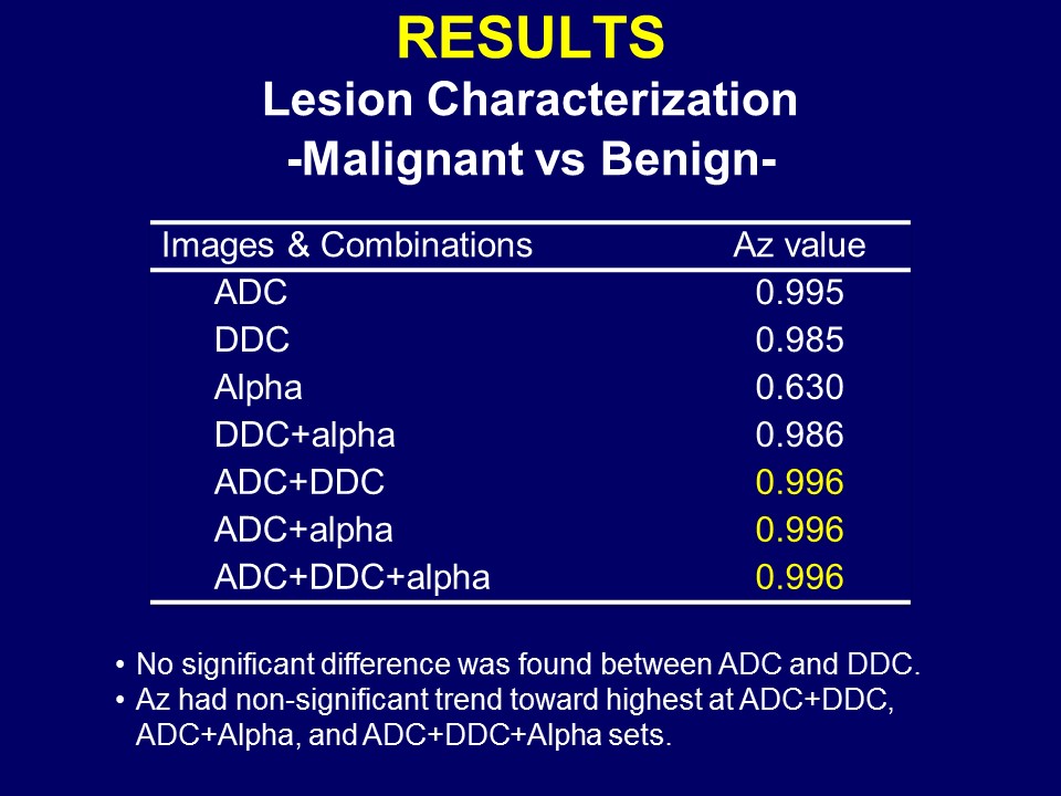
Figure 4. Lesion
Characterization
-Malignant vs Benign-
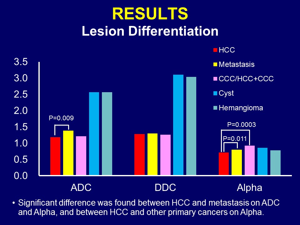
Figure 5. Malignant Lesion
Differentiation
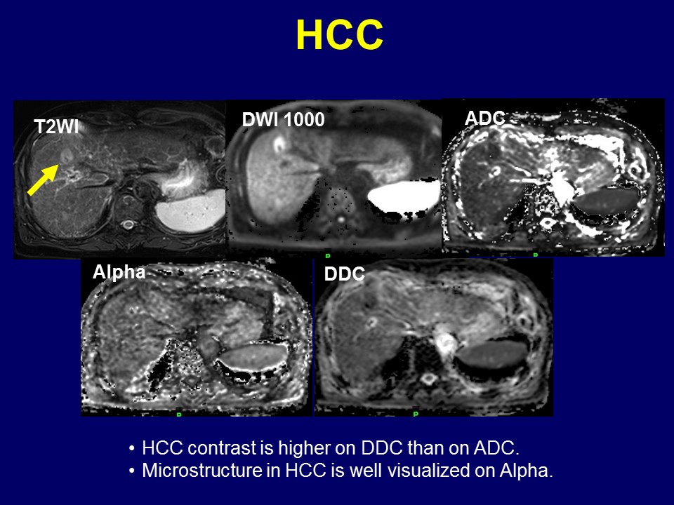
Figure 6. A Case of Hepatocellular Carcinoma
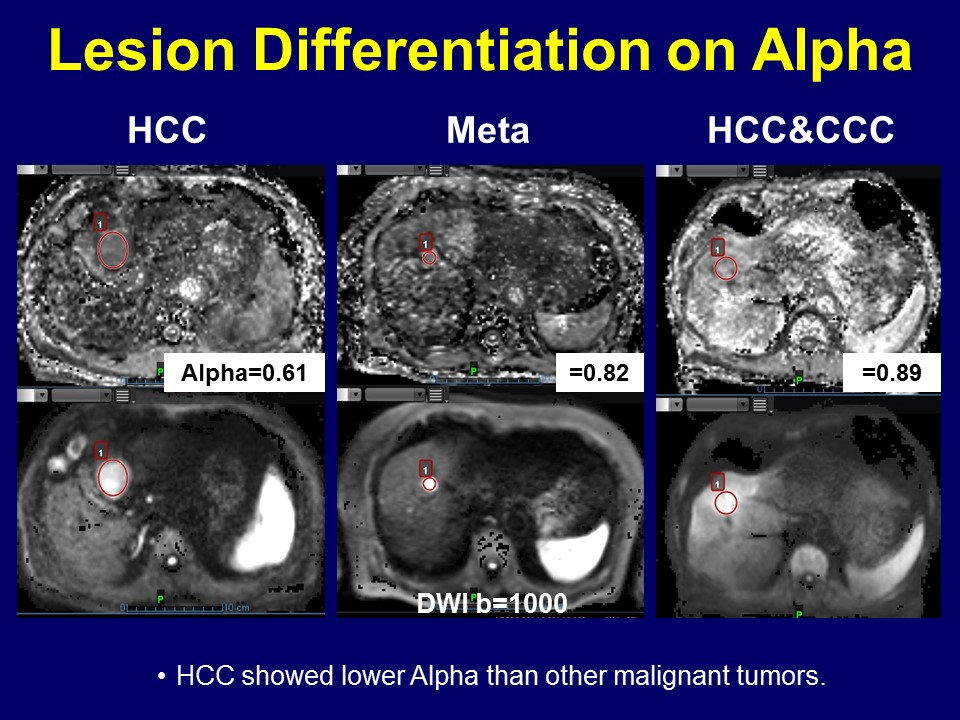
Figure 7. Malignant Lesion Differentiation on Alpha