2615
Optimizing diffusion-weighted imaging of thyroid gland using dedicated surface coil
Yunfei Wang1, Chaofan Zhu1, Dehong Luo1, Long Qian2, Yayuan Geng3, Wenming Deng1, and Zhou Liu1
1Radiology, National Cancer Center/National Clinical Research Center for Cancer/Cancer Hospital & Shenzhen Hospi, Shenzhen, China, 2MR Research, GE Healthcare, Beijing, China, 3Huiying Medical Technology Co., Ltd., Beijing, China
1Radiology, National Cancer Center/National Clinical Research Center for Cancer/Cancer Hospital & Shenzhen Hospi, Shenzhen, China, 2MR Research, GE Healthcare, Beijing, China, 3Huiying Medical Technology Co., Ltd., Beijing, China
Synopsis
It is challenging to perform diffusion-weighted imaging (DWI) on thyroid gland with high quality due to susceptibility artifacts caused by complex structure compositions, and physiological motion artifacts and low signal-to-noise ratio (SNR) using conventional head and neck joint coil. We used a histogram-based approach to evaluate the images of the small field-of-view (FOV) DWI (Focus, GE) and DWI with short-tau inversion-recovery (STIR) fat suppression using a state-of-the-art surface-coil specifically designed for thyroid gland. We found significantly different level and considerably inhomogeneity in signal intensity between right and left lobe of thyroid gland and between two DWIs.
Introduction
It is estimated that each individual has a 10% probability for developing thyroid nodules in their lifetime and 4.0-6.5% of detected thyroid nodules are malignant [1], which poses a huge challenge to clinicians in managing such vast number of thyroid nodules. Multiple studies have shown that diffusion weighted imaging (DWI) has great potential in differentiating benign and malignant thyroid nodules [2], which is however usually of inferior quality in imaging thyroid gland due to susceptibility artifacts caused by heterogeneous components of head and neck region, physiological motions including breathing, swallowing, coughing and joint movements and low signal-to-noise ratio using conventional head and neck joint coil [3]. The improvement of DWI image quality may come from better designed receiver coil and optimized acquisition sequence. In this study, a specifically designed surface coil is used in combination of different imaging strategies to achieve improved DWI image quality.Materials and Methods
This prospective study was approved by local institutional review board, and 36 healthy volunteers without any thyroid diseases were enrolled (male: female = 11: 25, 29.7 ± 6.6 years old) in the study. All participants underwent MR scan on a 3.0 T whole-body MRI scanner (Discovery MR 750, GE, US) equipped with a surface-coil (TH80-3T, Suzhou Medcoil Heathcare Co., Ltd, China) specifically designed for thyroid gland (Figure 1). Consistent placement of thyroid coil was checked for different subjects. Reduced field of view DWI (TR/TE: 3179/74-186ms, matrix size of 256×256, FOV: 14×4.2cm, slice thickness: 4 mm, b value of 0, and 1000) and DW-EPI DWI with short-tau inversion-recovery (STIR) fat suppression (TR/TE: 2857/75-174ms, matrix size of 256×256, FOV: 24×12cm, b value of 0, and 1000) were scanned. The image quality was assessed based on histogram-based features Firstly, on the radcloud platform (Huiying Medical Technology Co., Ltd, Beijing, China), two experienced radiologists independently manually segmented respective volumes of interest (VOIs) on the right and left lobe of thyroid gland on the contiguous slices of diffusion-weighted images; then 5 histogram-based features including mean, kurtosis, entropy, variance and skewness were extracted. To test reproducibility, intraclass correlation (ICC) was used to analyze the 4 measurements from right lobe on FOCUS DWI, left lobe on FOCUS DWI, right lobe on STIR-based DWI and left lobe on STIR-based DWI. Given the heath nature of all participants, quantitative measurements obtained from right and left lobe from the same DWI sequence and same subject on different DWIs were compared using t-test and Pearson’s correlation to test the image homogeneity with P < 0.01 indicating statistically significant difference.Results
Overall, well-defined boundary of thyroid gland was obtained using both FOCUS DWI and STIR-based DWI (Figure 2). Excellent inter-rater agreement supported by the ICC values was obtained: right lobe on FOCUS DWI (0.969, 95% confidence interval (CI): 0.677-0.991), left lobe on FOCUS DWI (0.947, 95%CI: 0.880-0.975), right lobe on STIR-based DWI (0.931, 95%CI: 0.580-0.978) and left lobe on STIR-based DWI (0.939, 95%CI: 0.877-0.970). The extracted measurements of both lobes using different DWI methods are summarized in Table 1. It can be seen that despite highly correlated with each other, the mean signal intensity of right lobe of thyroid gland was considerably significantly higher than the contralateral left lobe, both for FOCUS DWI and STIR-based DWI (both P < 0.001). This bias can also be visualized in Figure 2, and was a result of the non-uniform receiver sensitivity of the surface coil. On the other hand, the histogram-based measurements revealed the two lobes differed in variance and entropy but not in kurtosis nor skewness, indicating similar skewness and symmetry of the measurements but not the distributions (Figure 3). Similarly, for the same lobe, the intensity distribution of two DWI differs in variance and entropy, which demonstrates the heterogeneous signal intensity distribution between two DWIs (Figure 3).Discussion and Conclusions
Imaging of thyroid gland is challenging, especially when it comes to DWI. Improvement of DWI image quality hold promises for better diagnosis and treatment planning. In this work, the use of a dedicated surface coil combined with different diffusion imaging strategy was investigated on healthy volunteers. Overall good image quality was obtained, however image inhomogeneity that led to difference in histogram-based analysis was resulted. The inhomogeneity was mostly attributed to the design of surface coil. Further studies with patients are needed to warren optimal DWI of thyroid gland.Acknowledgements
We would thank Long Qian from GE healthcare and Yayuan Geng from Huiying Medical Technology Co., Ltd. for their technical support.References
[1] Geanina P, Jacqueline J. Thyroid nodules. Med Clin North Am. 2012 Mar; 96(2): 329–349. doi: 10.1016/j.mcna.2012.02.002 [2] Chen L, Xu J, Bao J, et al Diffusion-weighted MRI in differentiating malignant from benign thyroid nodules: a meta-analysis BMJ Open 2016;6:e008413. doi: 10.1136/bmjopen-2015-008413 [3] Thoeny HC, De Keyzer F, King AD. Diffusion-weighted MR imaging in the head and neck. Radiology. 2012 Apr;263(1):19-32. doi: 10.1148/radiol.11101821.Figures
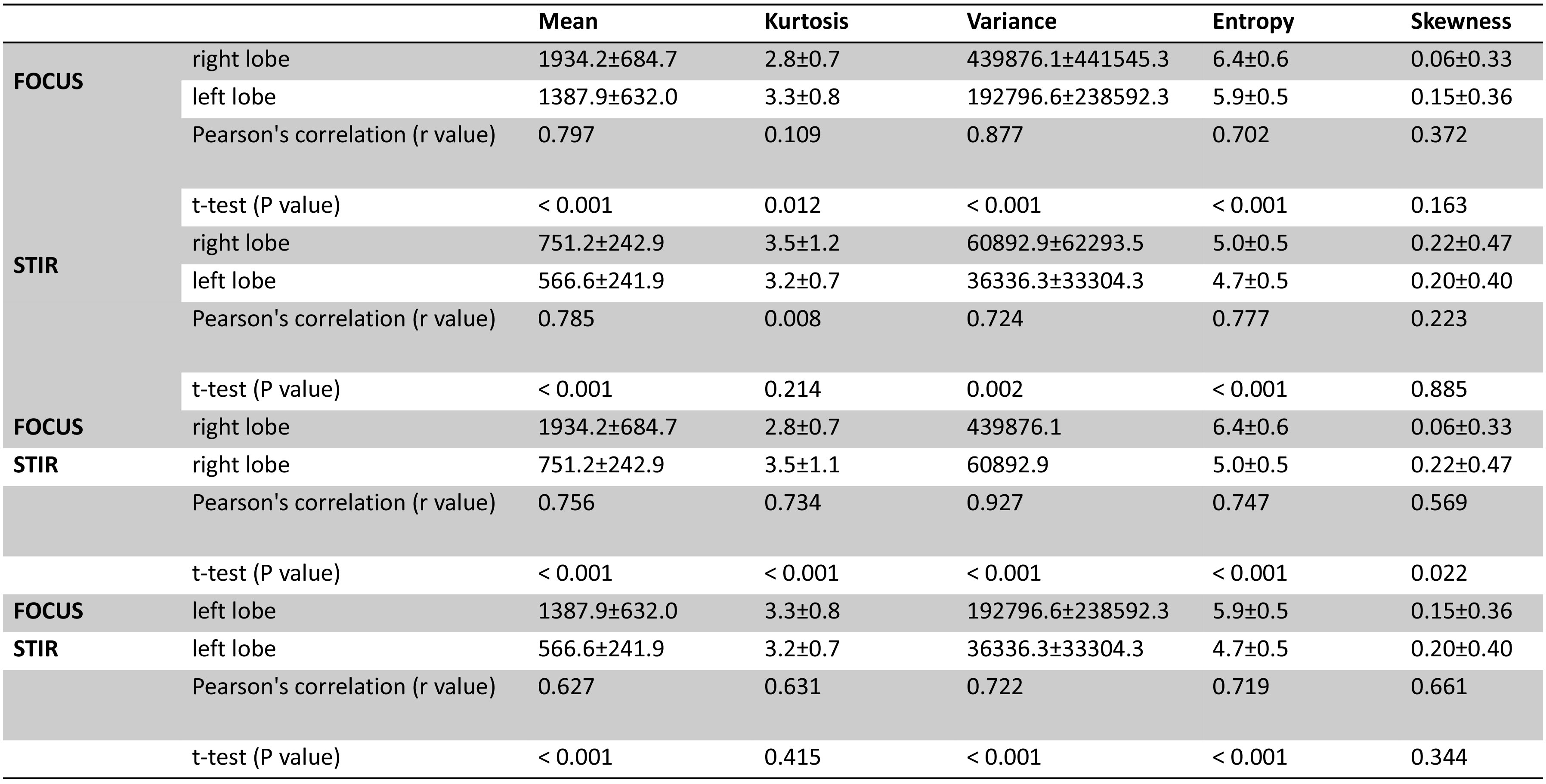
Table 1: histogram-based measurements of different
DWIs of left and right lobes of the thyroid
gland
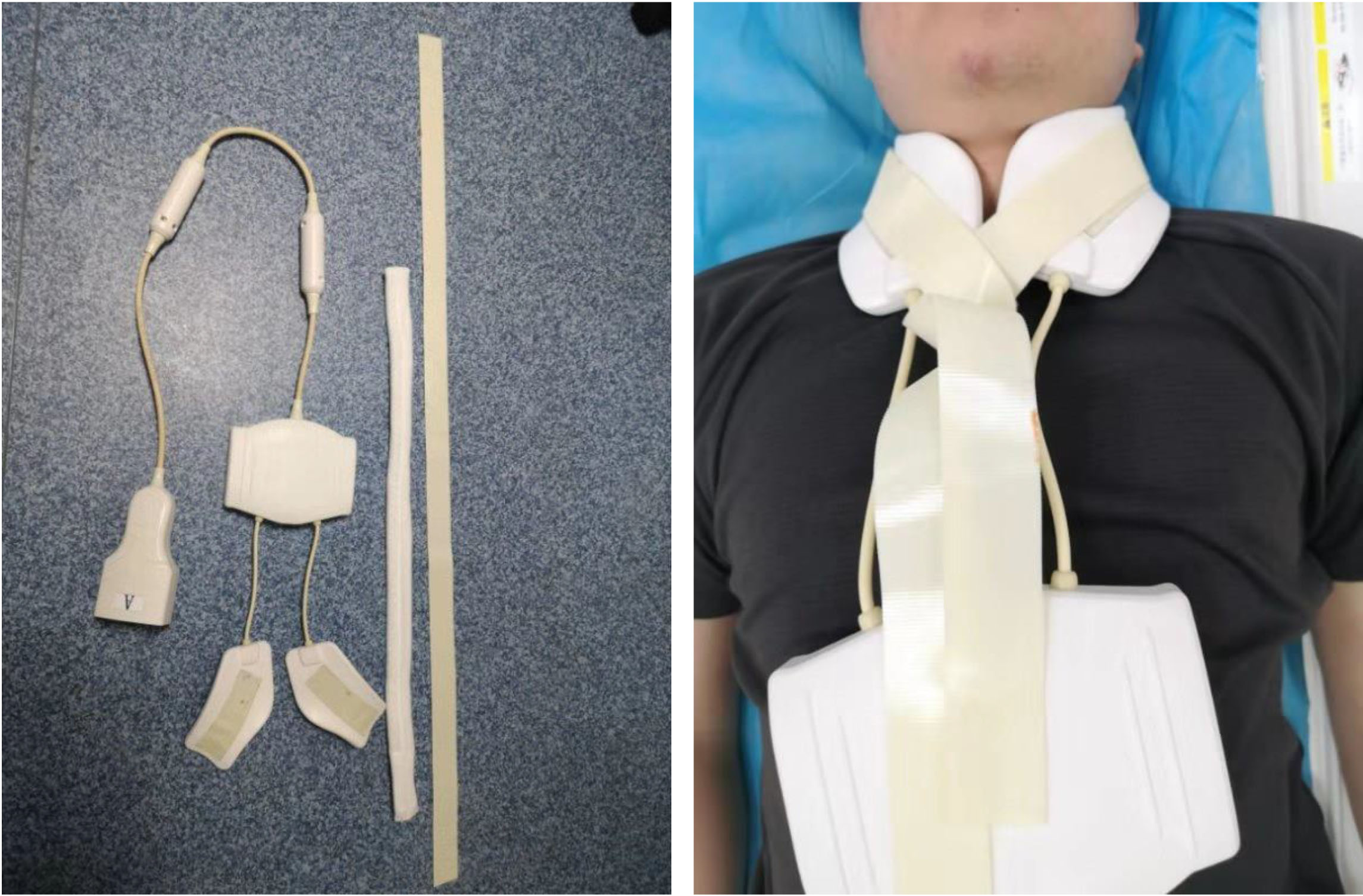
Figure 1. The surface-coil
specifically designed for thyroid gland and its placement.
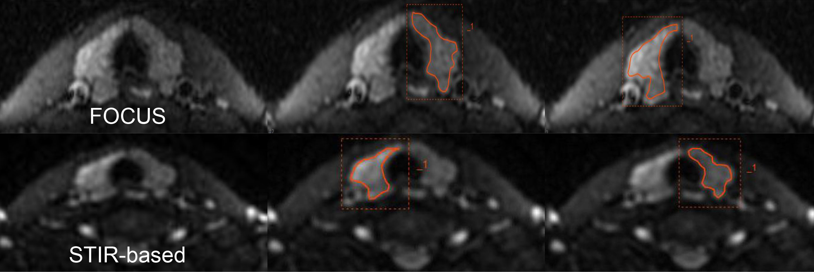
Figure 2. DWI images of a typical subject using
different acquisition methods along with the placement of VOIs.
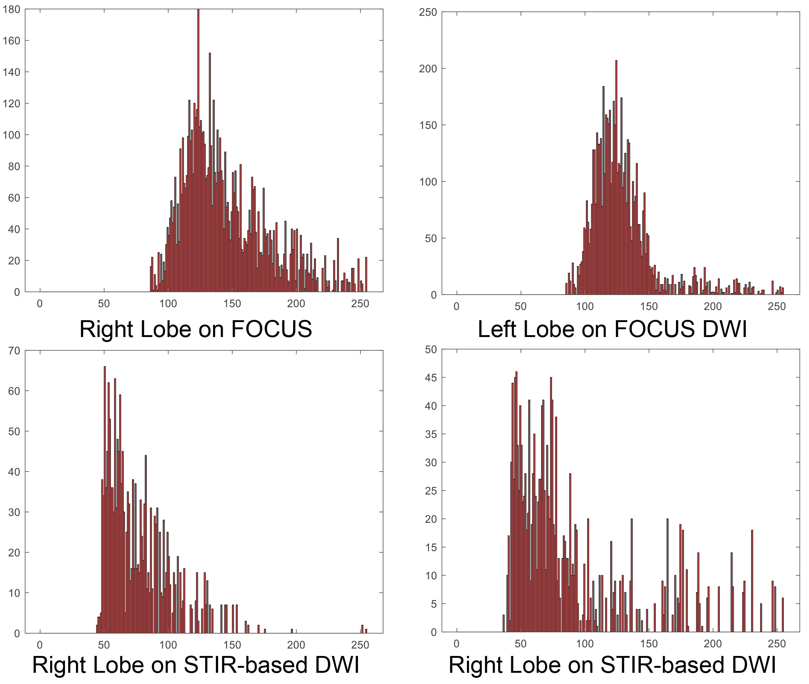
Figure 3. The histogram-based on a volume of interest (VOI) of
different lobes on different DWI sequences.