2608
Motion-Robust, Reduced-Distortion Diffusion MRI of the Liver with Optimized Motion Compensated Waveforms and Multi-shot EPI1Radiology, University of Wisconsin Madison, Madison, WI, United States, 2Medical Physics, University of Wisconsin Madison, Madison, WI, United States, 3Applications and Workflow, GE Healthcare, Boston, MA, United States
Synopsis
Conventional liver diffusion MRI acquisitions suffer from several challenges including low spatial resolution, B0-induced distortions, and elastic motion-induced signal voids. In this work, motion-robust and distortion-corrected liver diffusion-weighted imaging (DWI) was enabled by combining optimized motion compensated diffusion waveforms with multi-shot EPI acquisitions. Quantitative validation of distortions and ADC measurements was performed in healthy volunteer to assess the robustness and reproducibility of the combined techniques, as well as the synergistic effect of motion-robust gradient waveform and multi-shot EPI acquisitions. Preliminary patient example demonstrated the feasibility in patients with metastatic liver lesions.
Introduction
Diffusion-weighted (DW)-MRI of the abdomen has various potential applications, including the detection, staging, and treatment monitoring of cancer. However, DW-MRI has substantial technical challenges1-3. Central among these challenges is the extreme sensitivity of DW-MRI to physiological motion, which leads to:1) Signal voids in tissues that experience non-rigid motion during the application of DW gradients (eg: the left lobe of the liver). These signal voids complicate diagnostic image interpretation and introduce bias in quantitative measures of diffusion (eg: ADC).
2) Unpredictable phase variations in the signal acquired from each excitation, which result in the widespread use of single shot echo-planar imaging (ssEPI) in DW-MRI. Unfortunately, ssEPI results in substantial image distortions and limited resolution in DW-MRI, particularly in complex magnetic susceptibility environments such as the abdomen.
In recent years, advanced techniques have been proposed to address these challenges. Optimized motion-robust DW gradients techniques6-8 have shown excellent promise to overcome the signal voids present in tissues that experience substantial motion, leading to reliable imaging and quantitative ADC mapping throughout the entire liver. In addition, phase-corrected multi-shot reconstruction techniques4 has been proposed for reduced-distortion, high-resolution multi-shot EPI (msEPI) based DW-MRI4-5. Further, we hypothesize that a synergistic combination may be feasible between motion-robust DW gradient waveforms (which may reduce the presence of motion-induced phase variations in the acquired data) and msEPI techniques for liver DW-MRI. In this study, we evaluated the robustness and reproducibility of combined motion-robust DW waveforms and msEPI acquisitions.
Methods
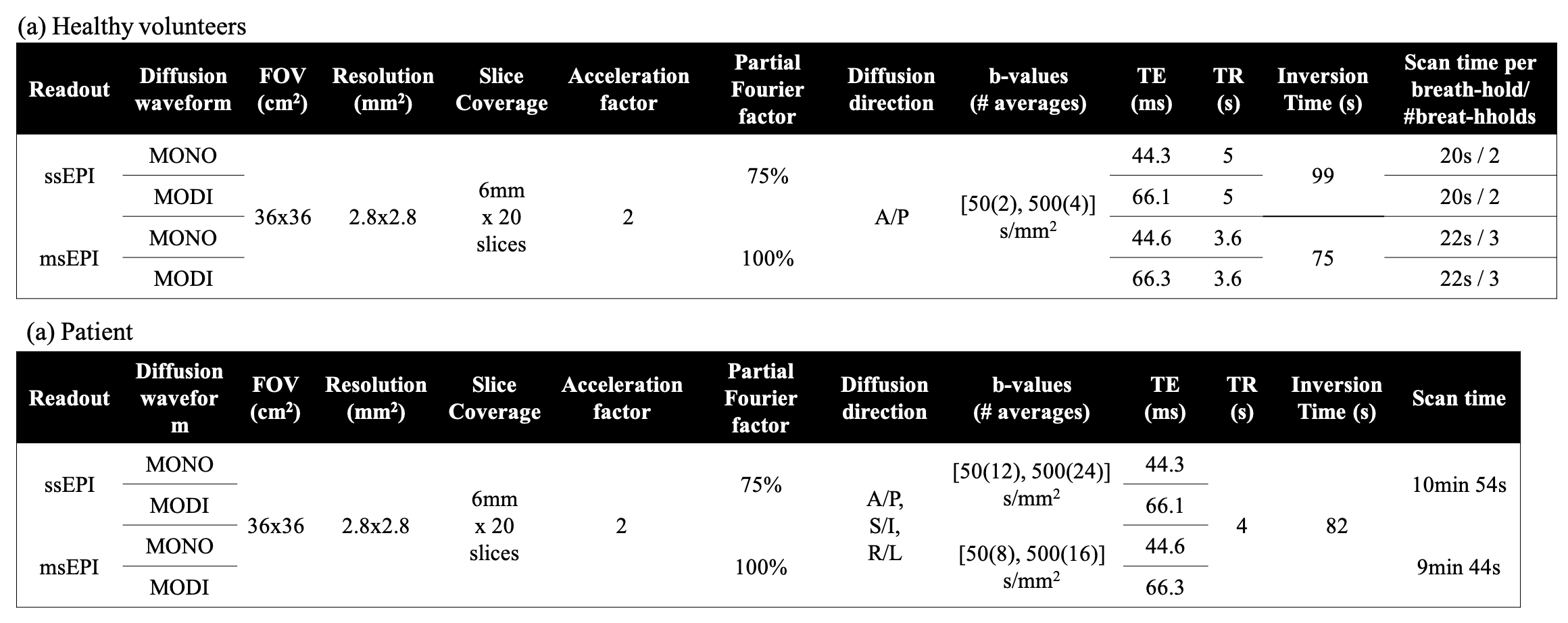
After IRB approval and informed written consent, eight healthy volunteers and one patient with liver metastases from primary pancreatic cancer were scanned on a 3T scanner (GE Signa Premier) with high-density receive array coils (AIR coil, GE Healthcare, Waukesha, WI). In comparison to the conventional monopolar (MONO) gradient waveform, M1-Optimized Diffusion Imaging (MODI) gradient waveform design8 was applied to achieve motion-robust diffusion waveforms with a small first-moment to suppress blood signal. For each DW waveform, two separate DW-MRI acquisitions using ssEPI and msEPI, respectively, were obtained. Detailed parameters are in Table.1. msEPI was obtained with two shots, each shot had acceleration factor=2 and reconstructed with a multiplexed sensitivity-encoding method4. DW-MRI acquisitions were performed during multiple breath-holding periods. Non-EPI T2-weighted (T2w) images were acquired with a 2D Fast Spin-Echo sequence as a reference of anatomic structure. The same spatial resolution and prescribed locations were used to match the DW-MRI acquisitions.ADC maps were calculated for all DW-MRI series. Co-localized ROIs were drawn on each ADC map in both the left and right liver lobes. The ADC in the non-motion-corrupted right liver lobe was used as reference ADC value2,6-8. Bland-Altman analysis was performed between the ADC measurements of the left and right lobe for each diffusion sequence to evaluate the consistency of ADC between the left and right lobes. Distortion level was assessed using the normalized cross-correlation coefficient (CCC)9, which quantifies the geometric alignment between the T2w reference and the low b-value DW images for each slice (Eq.1). Masks were selected from T2w references to remove the background noises. $$CCC=\frac{\sum_i{\sum_j{(T2_{ij}-\overline{T2})(DWI_{ij}-\overline{DWI})}}}{\sqrt{(\sum_i{\sum_j{(T2_{ij}-\overline{T2})^2}})(\sum_i{\sum_j{(DWI_{ij}-\overline{DWI})^2}})}}~~~Eq.1$$
Results
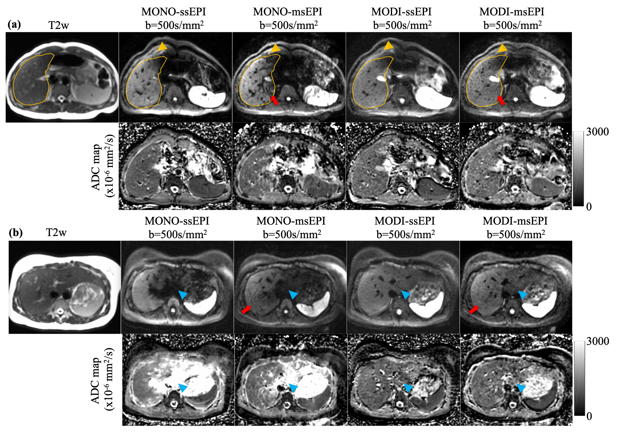
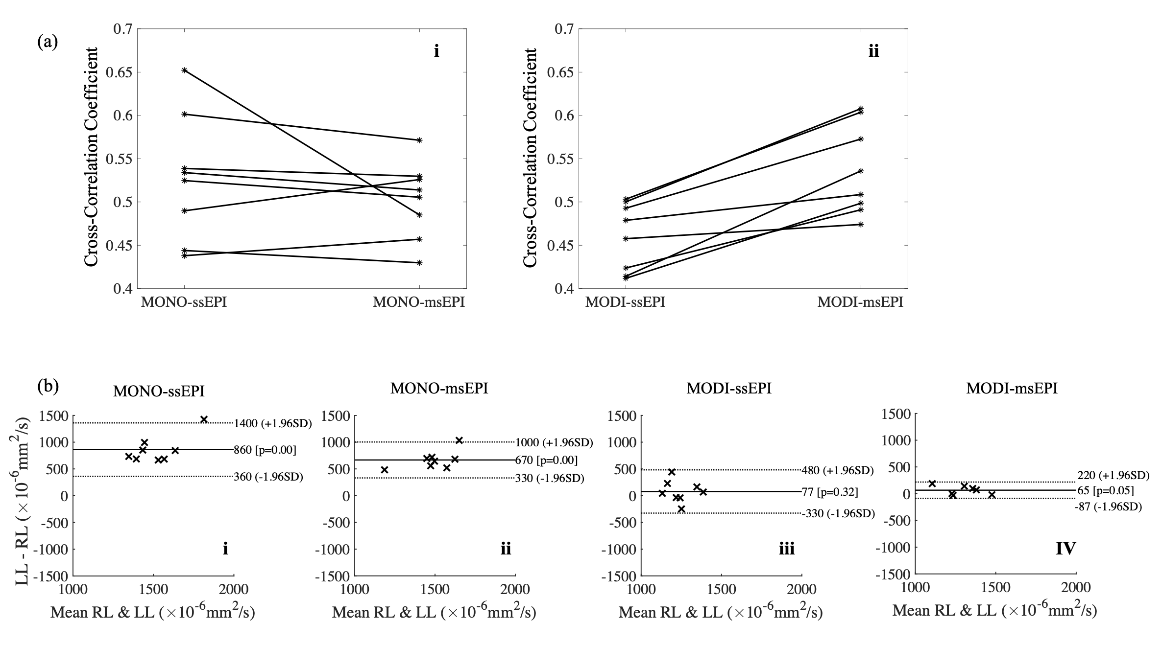
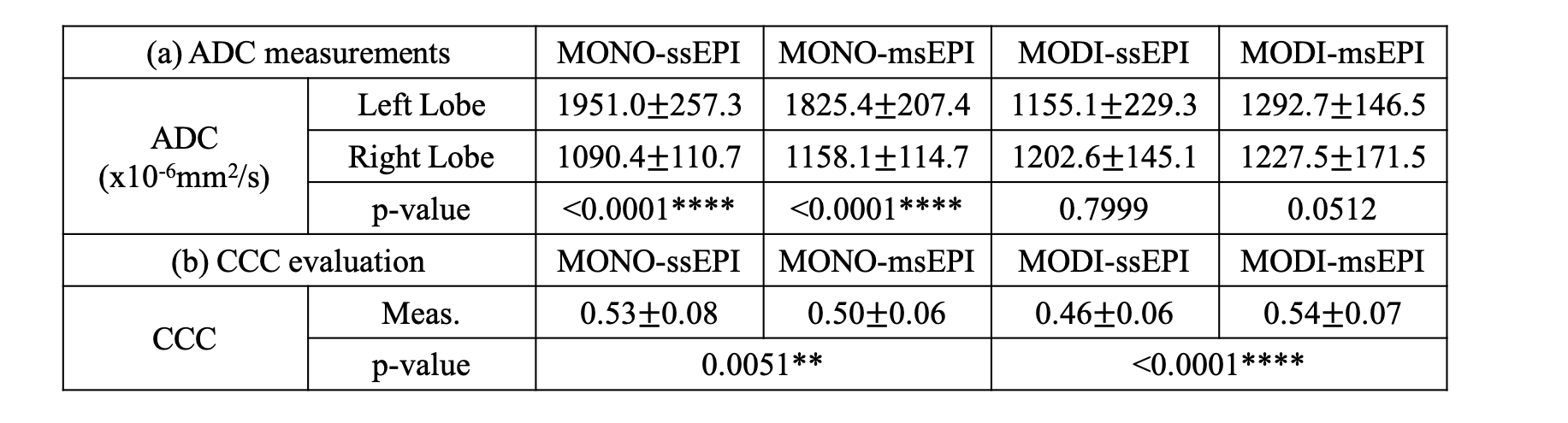
Fig.1 shows DW images from two healthy volunteers. The yellow curve in volunteer (a) depicts the contour of the liver from the T2w reference. The misalignment between the contour and the liver anatomy due to distortion in the DW images is indicated by yellow arrowheads. Images with msEPI acquisition have substantially reduced distortion artifacts. Blue arrowheads show the motion-induced signal voids and ADC bias in the left liver lobe, which was reduced by MODI acquisition. The worm-hole artifacts indicated by red arrows in the msEPI reconstruction with MONO acquisition were likely due to motion-induced rapid spatial phase variations, which were mitigated when combining msEPI with MODI.CCC comparison in Fig.2(a) illustrates the synergy between motion-robust diffusion waveform and msEPI acquisition. The alignment between DW images from MONO-ssEPI and the T2w reference is higher than MONO-msEPI (p=0.0051) even though the visually observed distortion artifact is largely reduced in MONO-msEPI (Fig.1). This is likely due to the substantial signal voids in the left liver lobe and the worm-hole artifacts. With MODI waveform (ii), msEPI has significant improvement (p<0.0001) in terms of alignment, which indicates significantly reduced distortion.
Bland-Altman analysis in Fig.2(b) demonstrates reduced ADC bias of the left liver lobe due to the motion-robust MODI acquisition. The ADC values in the left liver lobe of MONO-ssEPI and MONO-msEPI have significant bias compared to the right lobe. With MODI acquisitions, no significant bias was observed in both ssEPI and msEPI acquisitions.
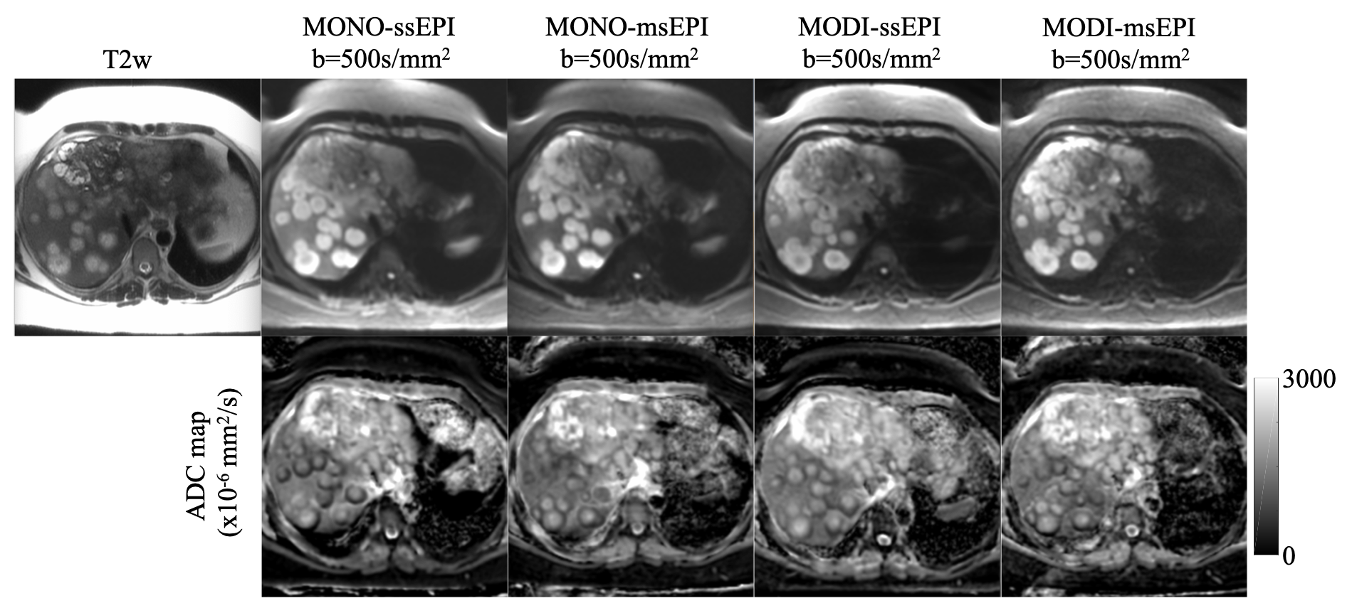
Finally, Fig.3 demonstrates the clinical feasibility of MODI-msEPI, which enabled high image quality with reduced distortions, in a patient with multiple liver metastases.
Discussion
In this study, we have investigated the feasibility and reproducibility of motion-robust and distortion-corrected DW-MRI by combining optimized motion-compensated diffusion waveforms (i.e. MODI) and msEPI acquisition. Volunteer study showed promising image quality with reduced ADC bias in the left lobe as well as reduced distortion. Importantly, this work demonstrated the potential synergy of combining MODI and msEPI techniques. Future studies will be focused on further evaluation in clinical patients.Conclusion
The synergetic effects of combined motion-compensated diffusion waveforms and msEPI acquisitions can enable reproducible and robust liver DWI with motion-robustness and reduced distortion.Acknowledgements
The authors acknowledge research support from GE Healthcare.References
[1] Naganawa, S., Kawai, H., Fukatsu, H., Sakurai, Y., AOKI, I., MIURA, S., MIMURA, T., KANAZAWA, H. and ISHIGAKI, T., 2005. Diffusion-weighted imaging of the liver: technical challenges and prospects for the future. Magnetic Resonance in Medical Sciences, 4(4), pp.175-186.
[2] Murphy, P., Wolfson, T., Gamst, A., Sirlin, C. and Bydder, M., 2013. Error model for reduction of cardiac and respiratory motion effects in quantitative liver DW‐MRI. Magnetic resonance in medicine, 70(5), pp.1460-1469.
[3] Kwee, T.C., Takahara, T., Niwa, T., Ivancevic, M.K., Herigault, G., Van Cauteren, M. and Luijten, P.R., 2009. Influence of cardiac motion on diffusion-weighted magnetic resonance imaging of the liver. Magnetic Resonance Materials in Physics, Biology and Medicine, 22(5), pp.319-325.
[4] Chen, N.K., Guidon, A., Chang, H.C. and Song, A.W., 2013. A robust multi-shot scan strategy for high-resolution diffusion weighted MRI enabled by multiplexed sensitivity-encoding (MUSE). Neuroimage, 72, pp.41-47.
[5] Zhang, Y., Holmes, J., Rabanillo, I., Guidon, A., Wells, S. and Hernando, D., 2018. Quantitative diffusion MRI using reduced field-of-view and multi-shot acquisition techniques: Validation in phantoms and prostate imaging. Magnetic resonance imaging, 51, pp.173-181.
[6] Aliotta, E., Wu, H.H. and Ennis, D.B., 2017. Convex optimized diffusion encoding (CODE) gradient waveforms for minimum echo time and bulk motion–compensated diffusion‐weighted MRI. Magnetic resonance in medicine, 77(2), pp.717-729.
[7] Peña‐Nogales, Ó., Zhang, Y., Wang, X., de Luis‐Garcia, R., Aja‐Fernández, S., Holmes, J. H., & Hernando, D. (2019). Optimized Diffusion‐Weighting Gradient Waveform Design (ODGD) formulation for motion compensation and concomitant gradient nulling. Magnetic resonance in medicine, 81(2), 989-1003
[8] Zhang Y, Peña‐Nogales Ó, Holmes JH, Hernando D. Motion‐robust and blood‐suppressed M1‐optimized diffusion MR imaging of the liver. Magnetic resonance in medicine. 2019 Jul;82(1):302-11.
[9] Hancu I, Lee SK, Hulsey K, et al. Distortion correction in diffusion-weighted imaging of the breast: Performance assessment of prospective, retrospective, and combined (prospective + retrospective) approaches. Magn Reson Med. 2017;78(1):247-253. doi:10.1002/mrm.26328
Figures