2600
Accelerated Whole-body Imaging with Uniform Fat-Suppression and Deep-Learning Reconstruction1Radiology, University of Wisconsin Madison, Madison, WI, United States, 2Global MR Applications & Workflow, GE Healthcare, Waukesha, WI, United States, 3Global MR Applications & Workflow, GE Healthcare, Calgary, AB, Canada, 4Global MR Applications & Workflow, GE Healthcare, Houston, TX, United States
Synopsis
We present a single-shot fast-spin-echo pulse-sequence (SSFSE) with short-tau inversion recovery (STIR) and deep-learning (DL) image reconstruction that provides whole-body images in less than 3 minutes, achieving uniform fat suppression, detailed delineation of the anatomy, and favorable signal-to-noise (SNR). This was achieved through the reduction of echo-train-length by increasing the acceleration to a factor of 4x, hence substantially improving image quality through blurring reduction in the phase encoding direction, and ameliorating the subsequent SNR reductions by utilization of a DL reconstruction algorithm. This whole-body MRI technique can be used in the setting of various pathologies for both pediatrics and adults.
INTRODUCTION
There is an increasing clinical demand for whole-body (WB) MRI for a variety of purposes, including diagnosis/staging/follow-up of malignancies, as well as screening of high-risk patients [1]; one common example is detection of osseous lesions in the setting of breast cancer, renal cell carcinoma, and multiple myeloma. Utilization of WB-MRI acquisition techniques such as single-shot fast spin-echo (SSFSE) are necessary to improve patient throughput and reduce motion artifacts. However, single-shot techniques are subject to some challenges; for example, the possible induction of high RF power deposition exhibits objectionable blurring in the phase encoding direction due to vastly extended echo train lengths (ETLs). These effects can be mitigated to some extent by the use of variable flip angle schemes [2, 3], but still limit the overall performance and achievable ETL. The use of parallel acceleration techniques can reduce the ETL by a factor of 2x or more, however with at the cost of substantially-reduced signal-to-noise (SNR). Recently, deep learning-based reconstruction approaches have demonstrated promise to recover image SNR. Therefore, the purpose of this work is to study highly-accelerated SSFSE for WB-MRI using a deep learning-based reconstruction.METHODS
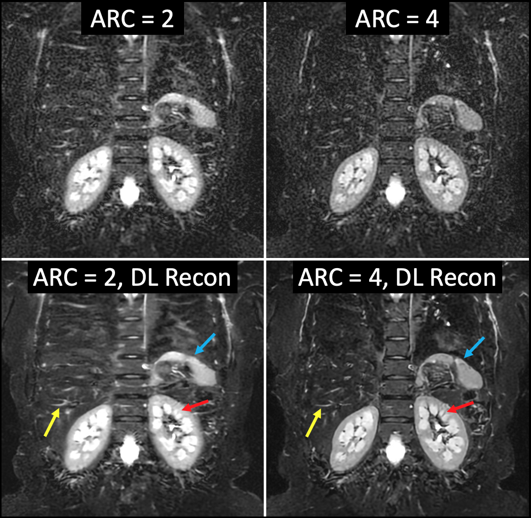
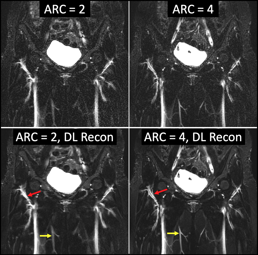
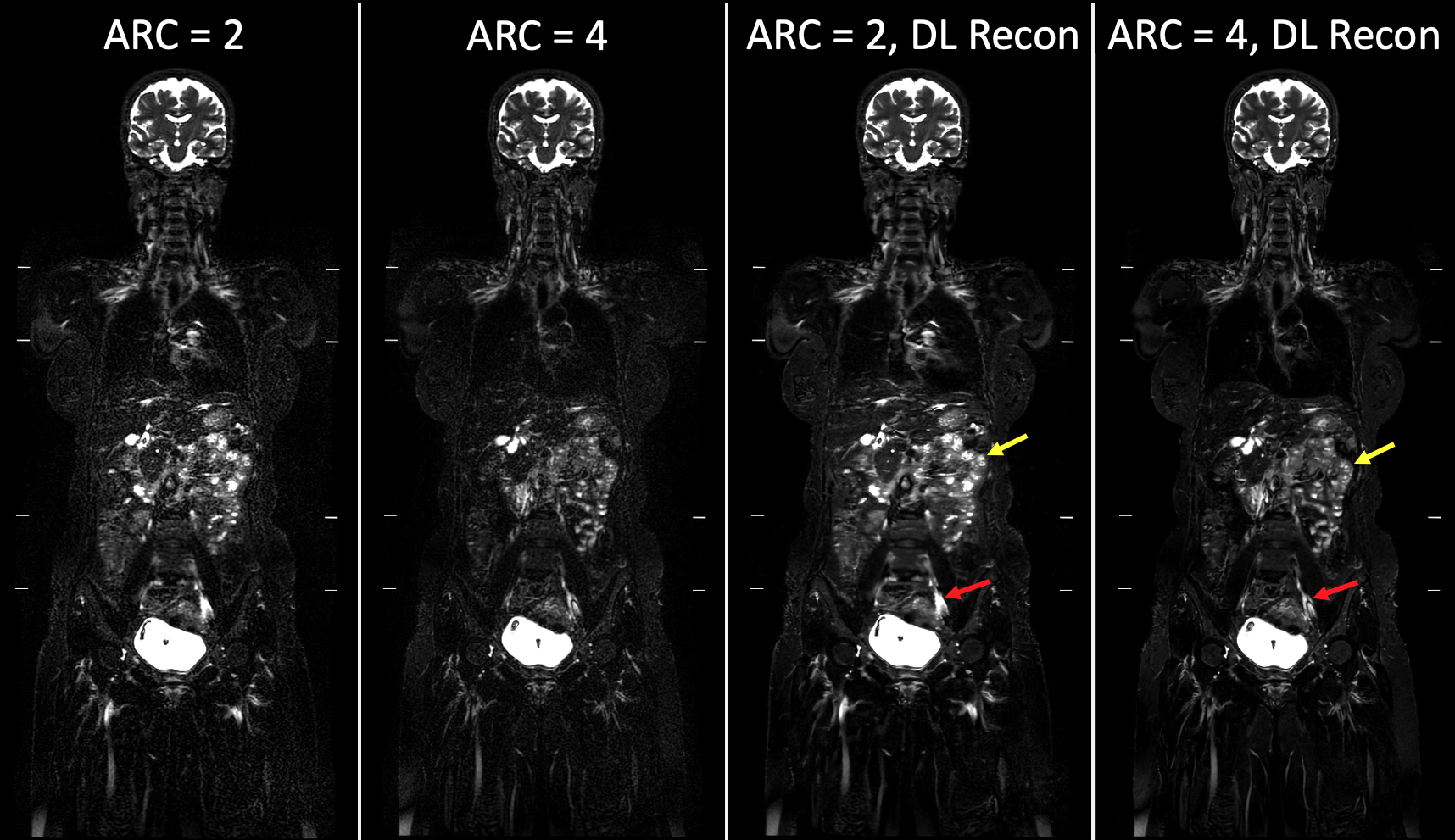
Imaging studies were performed on a 1.5T MRI scanner (Artist, GE Healthcare, Waukesha, USA), using a whole-body coil suite (head/neck unit, AIR anterior array, posterior array; GE Healthcare). A SSFSE-STIR pulse sequence was optimized in the coronal plane with the following parameters: TR = 571 msec, TE = 96 msec, TI = 150 msec, ETL = 107 matrix = 288 x 224 (frequency x phase), voxel size = 1.5 x 2 x 4 mm, bandwidth = 83.3 kHz, craniocaudal coverage per station = 44 cm, oversampling = 1.2, tailored RF pulse, scan time per station = 24 sec. Imaging was repeated with increasing acceleration (ARC = 4) to reduce blurring; TR was adjusted to 476 msec, and ETL decreased to 82, yielding a scan time of 20 sec per station. Three stations were scanned to cover the anatomy from skull vertex to thighs. Images were reconstructed using both the conventional as well as a prototype deep-learning (DL Recon, GE Healthcare) reconstruction algorithms. Images at different stations were fused to generate a single set of whole-body images.RESULTS
An increase in image acquisition acceleration (from 2x to 4x) resulted in markedly reduced blurring, evidenced by a favorable delineation of the vasculature, nerves, and the contour of various organs, such as the spine, liver, spleen and kidneys in an example case (Figures 1-3). However, this improvement was observed at the cost of increased image noise and a less favorable SNR using conventional accelerated reconstructions. The implementation of the DL Recon algorithm achieved a substantially appreciable reduction in noise level without the loss of fine detail in the image; in fact, some structures, such as the liver vasculature, are better evaluated after the implementation of the DL reconstruction algorithm.DISCUSSION/CONCLUSIONS
Development of a rapid whole-body MRI pulse sequence with robust fat suppression and the ability for tumor detection is an area that has been challenging due to the limitations of the conventional pulse sequences and image reconstruction algorithms; often SNR and image detail are compromised by limits on image acquisition time. The presented SSFSE-STIR acquisition with DL reconstruction shows great promise, as it provides whole-body images in less than 3 minutes, with uniform fat suppression, detailed delineation of the anatomy, and favorable SNR. The reduction of ETL, enabled by increasing the acceleration to a factor of 4x, greatly improves image quality by reducing the blurring in the phase encoding direction for which SNR reductions due to acceleration are ameliorated by the DL reconstruction algorithm. This sequence can be used as an excellent complement to PET in the setting of PET/MRI, or in whole-body MRI without PET in the setting of various pathologies, such as cancer or systemic inflammatory conditions. The rapid nature of this acquisition is highly applicable to both pediatric and adult patients.Acknowledgements
No acknowledgement found.References
1. Morone M, Bali MA, Tunariu N, et al. Whole-Body MRI: Current Applications in Oncology. AJR American journal of roentgenology 2017; 209:W336-W349
2. Busse RF, Hariharan H, Vu A, Brittain JH. Fast spin echo sequences with very long echo trains: design of variable refocusing flip angle schedules and generation of clinical T2 contrast. Magn Reson Med 2006; 55:1030-1037
3. Loening AM, Saranathan M, Ruangwattanapaisarn N, Litwiller DV, Shimakawa A, Vasanawala SS. Increased speed and image quality in single-shot fast spin echo imaging via variable refocusing flip angles. Journal of magnetic resonance imaging : JMRI 2015; 42:1747-1758
Figures